Fluorine »
PDB 3kqn-3l8s »
3l8s »
Fluorine in PDB 3l8s: Human P38 Map Kinase in Complex with Cp-547632
Enzymatic activity of Human P38 Map Kinase in Complex with Cp-547632
All present enzymatic activity of Human P38 Map Kinase in Complex with Cp-547632:
2.7.11.24;
2.7.11.24;
Protein crystallography data
The structure of Human P38 Map Kinase in Complex with Cp-547632, PDB code: 3l8s
was solved by
C.Gruetter,
J.R.Simard,
D.Rauh,
with X-Ray Crystallography technique. A brief refinement statistics is given in the table below:
| Resolution Low / High (Å) | 40.00 / 2.35 |
| Space group | P 21 21 21 |
| Cell size a, b, c (Å), α, β, γ (°) | 66.980, 69.100, 74.300, 90.00, 90.00, 90.00 |
| R / Rfree (%) | 23.9 / 30.7 |
Other elements in 3l8s:
The structure of Human P38 Map Kinase in Complex with Cp-547632 also contains other interesting chemical elements:
| Bromine | (Br) | 1 atom |
Fluorine Binding Sites:
The binding sites of Fluorine atom in the Human P38 Map Kinase in Complex with Cp-547632
(pdb code 3l8s). This binding sites where shown within
5.0 Angstroms radius around Fluorine atom.
In total 2 binding sites of Fluorine where determined in the Human P38 Map Kinase in Complex with Cp-547632, PDB code: 3l8s:
Jump to Fluorine binding site number: 1; 2;
In total 2 binding sites of Fluorine where determined in the Human P38 Map Kinase in Complex with Cp-547632, PDB code: 3l8s:
Jump to Fluorine binding site number: 1; 2;
Fluorine binding site 1 out of 2 in 3l8s
Go back to
Fluorine binding site 1 out
of 2 in the Human P38 Map Kinase in Complex with Cp-547632
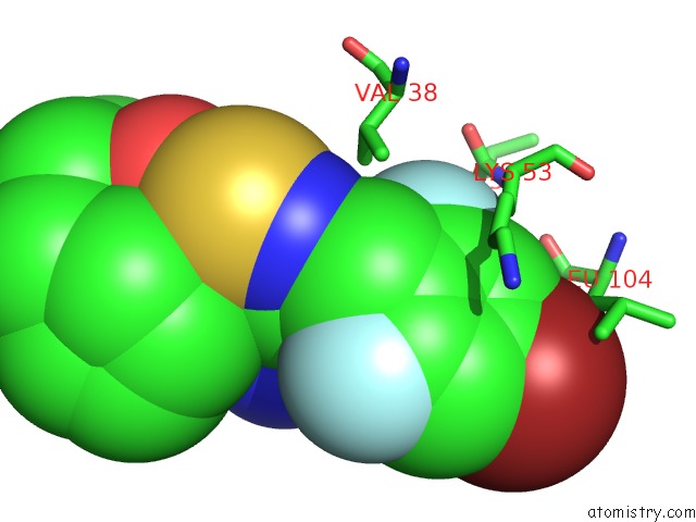
Mono view
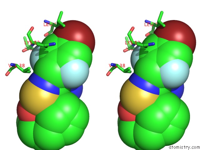
Stereo pair view
Mono view
Stereo pair view
A full contact list of Fluorine with other atoms in the F binding
site number 1 of Human P38 Map Kinase in Complex with Cp-547632 within 5.0Å range:
|
Fluorine binding site 2 out of 2 in 3l8s
Go back to
Fluorine binding site 2 out
of 2 in the Human P38 Map Kinase in Complex with Cp-547632
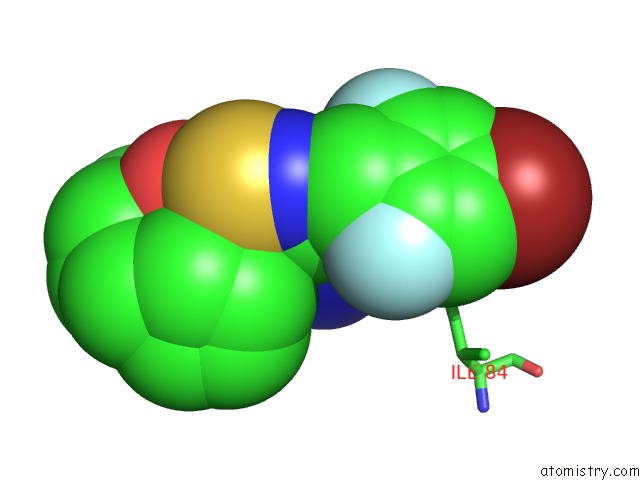
Mono view
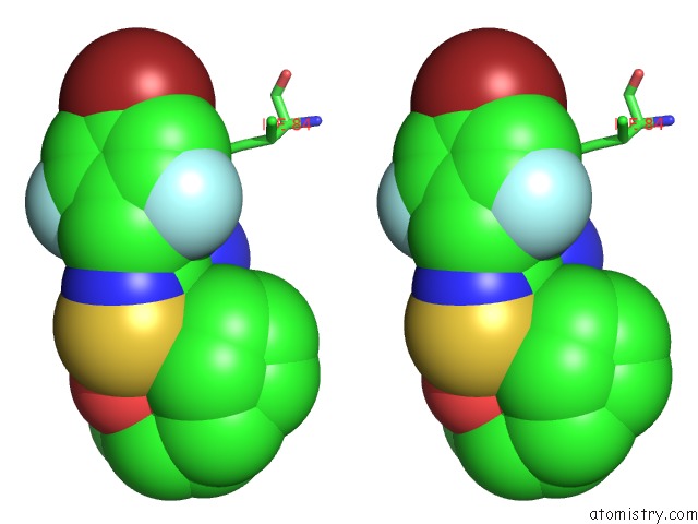
Stereo pair view
Mono view
Stereo pair view
A full contact list of Fluorine with other atoms in the F binding
site number 2 of Human P38 Map Kinase in Complex with Cp-547632 within 5.0Å range:
|
Reference:
J.R.Simard,
M.Getlik,
C.Grutter,
R.Schneider,
S.Wulfert,
D.Rauh.
Fluorophore Labeling of the Glycine-Rich Loop As A Method of Identifying Inhibitors That Bind to Active and Inactive Kinase Conformations. J.Am.Chem.Soc. V. 132 4152 2010.
ISSN: ISSN 0002-7863
PubMed: 20201574
DOI: 10.1021/JA908083E
Page generated: Wed Jul 31 20:24:03 2024
ISSN: ISSN 0002-7863
PubMed: 20201574
DOI: 10.1021/JA908083E
Last articles
Zn in 9J0NZn in 9J0O
Zn in 9J0P
Zn in 9FJX
Zn in 9EKB
Zn in 9C0F
Zn in 9CAH
Zn in 9CH0
Zn in 9CH3
Zn in 9CH1