Fluorine »
PDB 3l8v-3lxp »
3l9h »
Fluorine in PDB 3l9h: X-Ray Structure of Mitotic Kinesin-5 (Ksp, KIF11, EG5)in Complex with the Hexahydro-2H-Pyrano[3,2-C]Quinoline Emd 534085
Protein crystallography data
The structure of X-Ray Structure of Mitotic Kinesin-5 (Ksp, KIF11, EG5)in Complex with the Hexahydro-2H-Pyrano[3,2-C]Quinoline Emd 534085, PDB code: 3l9h
was solved by
T.Knoechel,
with X-Ray Crystallography technique. A brief refinement statistics is given in the table below:
| Resolution Low / High (Å) | 30.00 / 2.00 |
| Space group | C 1 2 1 |
| Cell size a, b, c (Å), α, β, γ (°) | 160.950, 79.718, 69.513, 90.00, 96.88, 90.00 |
| R / Rfree (%) | 23.4 / 25.8 |
Fluorine Binding Sites:
The binding sites of Fluorine atom in the X-Ray Structure of Mitotic Kinesin-5 (Ksp, KIF11, EG5)in Complex with the Hexahydro-2H-Pyrano[3,2-C]Quinoline Emd 534085
(pdb code 3l9h). This binding sites where shown within
5.0 Angstroms radius around Fluorine atom.
In total 6 binding sites of Fluorine where determined in the X-Ray Structure of Mitotic Kinesin-5 (Ksp, KIF11, EG5)in Complex with the Hexahydro-2H-Pyrano[3,2-C]Quinoline Emd 534085, PDB code: 3l9h:
Jump to Fluorine binding site number: 1; 2; 3; 4; 5; 6;
In total 6 binding sites of Fluorine where determined in the X-Ray Structure of Mitotic Kinesin-5 (Ksp, KIF11, EG5)in Complex with the Hexahydro-2H-Pyrano[3,2-C]Quinoline Emd 534085, PDB code: 3l9h:
Jump to Fluorine binding site number: 1; 2; 3; 4; 5; 6;
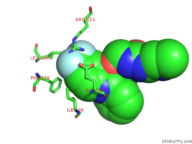
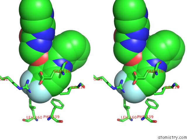
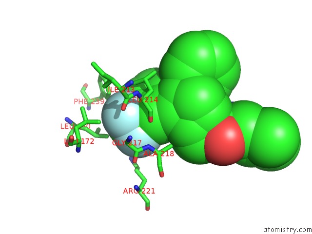
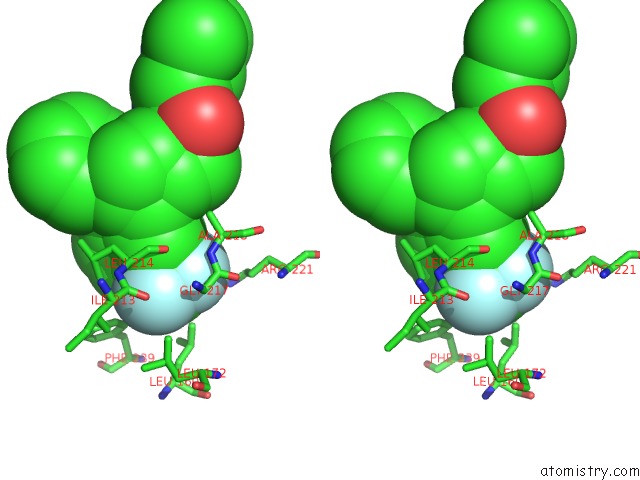
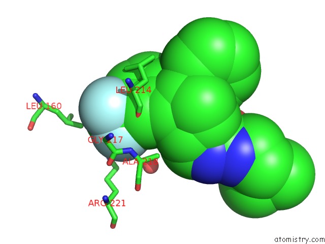
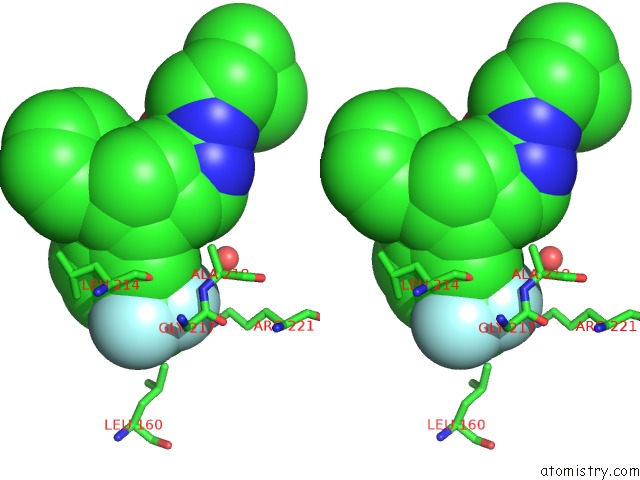
Fluorine binding site 1 out of 6 in 3l9h
Go back to
Fluorine binding site 1 out
of 6 in the X-Ray Structure of Mitotic Kinesin-5 (Ksp, KIF11, EG5)in Complex with the Hexahydro-2H-Pyrano[3,2-C]Quinoline Emd 534085
Mono view
Stereo pair view
Mono view
Stereo pair view
A full contact list of Fluorine with other atoms in the F binding
site number 1 of X-Ray Structure of Mitotic Kinesin-5 (Ksp, KIF11, EG5)in Complex with the Hexahydro-2H-Pyrano[3,2-C]Quinoline Emd 534085 within 5.0Å range:
|
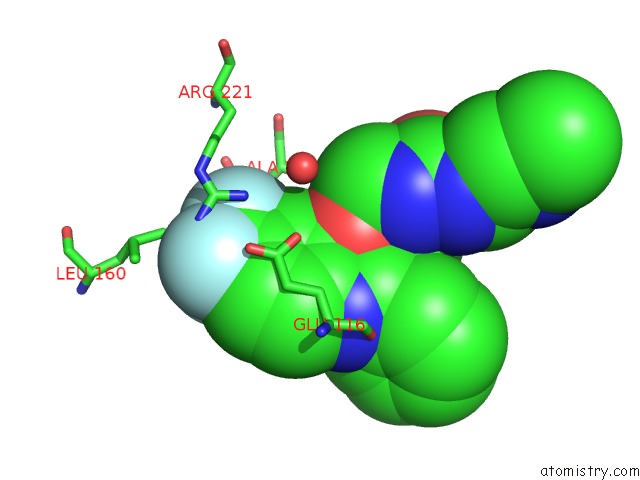
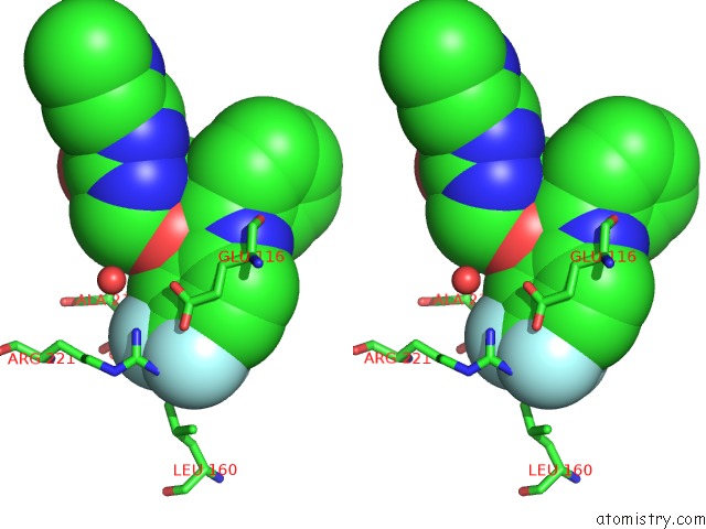
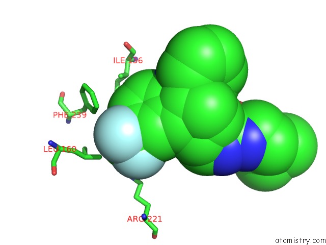
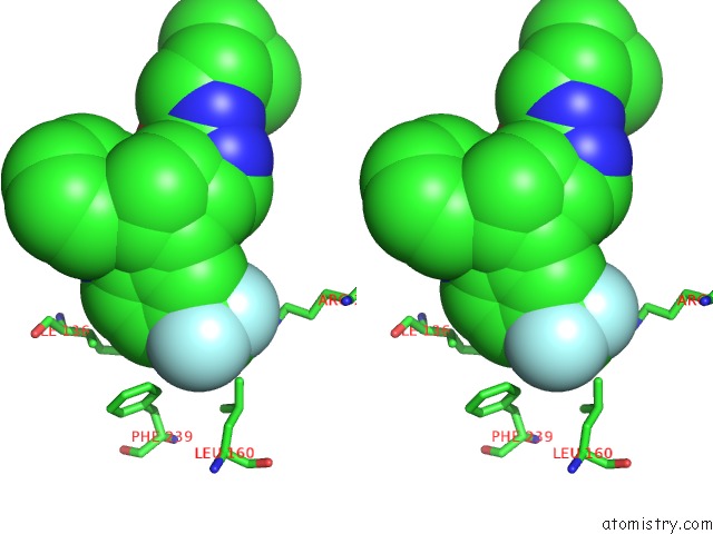
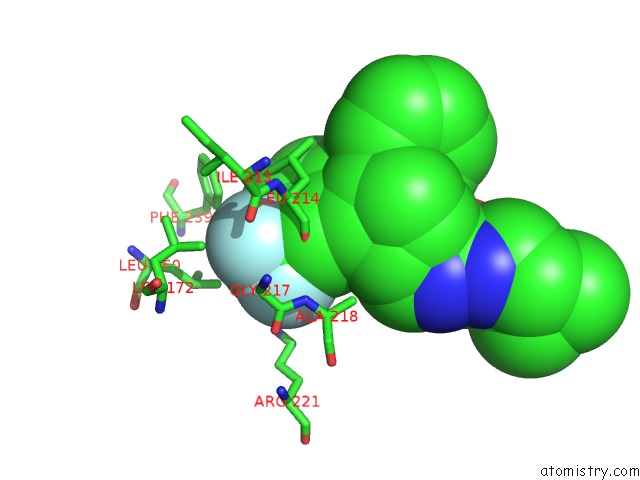
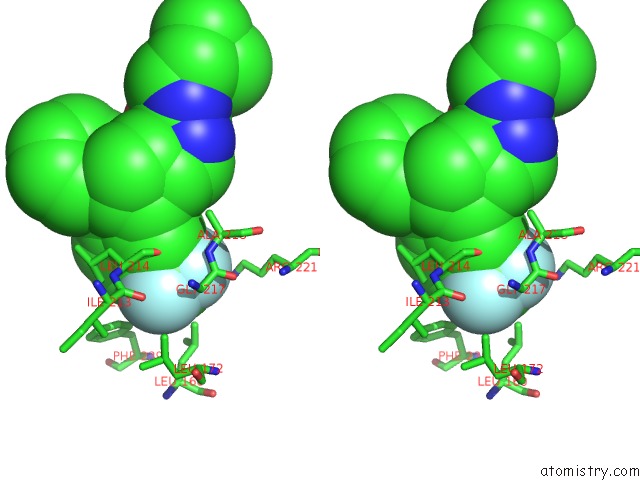
Fluorine binding site 2 out of 6 in 3l9h
Go back to
Fluorine binding site 2 out
of 6 in the X-Ray Structure of Mitotic Kinesin-5 (Ksp, KIF11, EG5)in Complex with the Hexahydro-2H-Pyrano[3,2-C]Quinoline Emd 534085
Mono view
Stereo pair view
Mono view
Stereo pair view
A full contact list of Fluorine with other atoms in the F binding
site number 2 of X-Ray Structure of Mitotic Kinesin-5 (Ksp, KIF11, EG5)in Complex with the Hexahydro-2H-Pyrano[3,2-C]Quinoline Emd 534085 within 5.0Å range:
|
Fluorine binding site 3 out of 6 in 3l9h
Go back to
Fluorine binding site 3 out
of 6 in the X-Ray Structure of Mitotic Kinesin-5 (Ksp, KIF11, EG5)in Complex with the Hexahydro-2H-Pyrano[3,2-C]Quinoline Emd 534085
Mono view
Stereo pair view
Mono view
Stereo pair view
A full contact list of Fluorine with other atoms in the F binding
site number 3 of X-Ray Structure of Mitotic Kinesin-5 (Ksp, KIF11, EG5)in Complex with the Hexahydro-2H-Pyrano[3,2-C]Quinoline Emd 534085 within 5.0Å range:
|
Fluorine binding site 4 out of 6 in 3l9h
Go back to
Fluorine binding site 4 out
of 6 in the X-Ray Structure of Mitotic Kinesin-5 (Ksp, KIF11, EG5)in Complex with the Hexahydro-2H-Pyrano[3,2-C]Quinoline Emd 534085
Mono view
Stereo pair view
Mono view
Stereo pair view
A full contact list of Fluorine with other atoms in the F binding
site number 4 of X-Ray Structure of Mitotic Kinesin-5 (Ksp, KIF11, EG5)in Complex with the Hexahydro-2H-Pyrano[3,2-C]Quinoline Emd 534085 within 5.0Å range:
|
Fluorine binding site 5 out of 6 in 3l9h
Go back to
Fluorine binding site 5 out
of 6 in the X-Ray Structure of Mitotic Kinesin-5 (Ksp, KIF11, EG5)in Complex with the Hexahydro-2H-Pyrano[3,2-C]Quinoline Emd 534085
Mono view
Stereo pair view
Mono view
Stereo pair view
A full contact list of Fluorine with other atoms in the F binding
site number 5 of X-Ray Structure of Mitotic Kinesin-5 (Ksp, KIF11, EG5)in Complex with the Hexahydro-2H-Pyrano[3,2-C]Quinoline Emd 534085 within 5.0Å range:
|
Fluorine binding site 6 out of 6 in 3l9h
Go back to
Fluorine binding site 6 out
of 6 in the X-Ray Structure of Mitotic Kinesin-5 (Ksp, KIF11, EG5)in Complex with the Hexahydro-2H-Pyrano[3,2-C]Quinoline Emd 534085
Mono view
Stereo pair view
Mono view
Stereo pair view
A full contact list of Fluorine with other atoms in the F binding
site number 6 of X-Ray Structure of Mitotic Kinesin-5 (Ksp, KIF11, EG5)in Complex with the Hexahydro-2H-Pyrano[3,2-C]Quinoline Emd 534085 within 5.0Å range:
|
Reference:
K.Schiemann,
D.Finsinger,
F.Zenke,
C.Amendt,
T.Knochel,
D.Bruge,
H.P.Buchstaller,
U.Emde,
W.Stahle,
S.Anzali.
The Discovery and Optimization of Hexahydro-2H-Pyrano[3,2-C]Quinolines (Hhpqs) As Potent and Selective Inhibitors of the Mitotic Kinesin-5. Bioorg.Med.Chem.Lett. V. 20 1491 2010.
ISSN: ISSN 0960-894X
PubMed: 20149654
DOI: 10.1016/J.BMCL.2010.01.110
Page generated: Wed Jul 31 20:27:54 2024
ISSN: ISSN 0960-894X
PubMed: 20149654
DOI: 10.1016/J.BMCL.2010.01.110
Last articles
Zn in 9MJ5Zn in 9HNW
Zn in 9G0L
Zn in 9FNE
Zn in 9DZN
Zn in 9E0I
Zn in 9D32
Zn in 9DAK
Zn in 8ZXC
Zn in 8ZUF