Fluorine in PDB 3lu6: Human Serum Albumin in Complex with Compound 1
Protein crystallography data
The structure of Human Serum Albumin in Complex with Compound 1, PDB code: 3lu6
was solved by
D.Buttar,
N.Colclough,
S.Gerhardt,
P.A.Macfaul,
S.D.Phillips,
A.Plowright,
P.Whittamore,
K.Tam,
K.Maskos,
S.Steinbacher,
H.Steuber,
with X-Ray Crystallography technique. A brief refinement statistics is given in the table below:
Resolution Low / High (Å)
|
55.64 /
2.70
|
Space group
|
P 1
|
Cell size a, b, c (Å), α, β, γ (°)
|
58.561,
59.868,
95.573,
74.51,
86.57,
74.47
|
R / Rfree (%)
|
23.6 /
27.5
|
Fluorine Binding Sites:
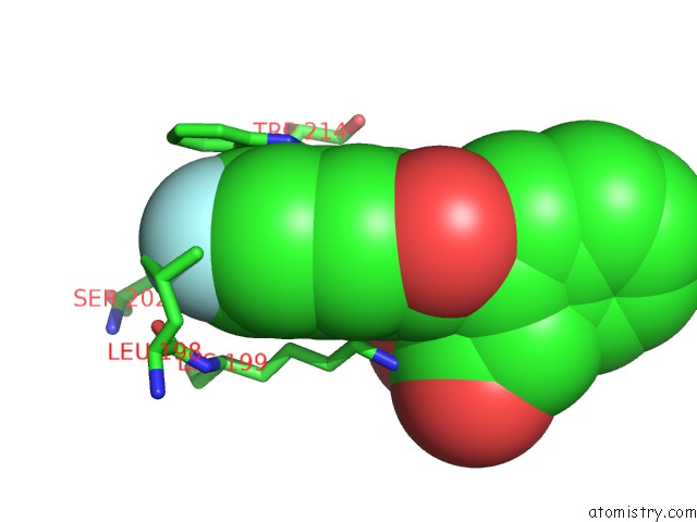
The binding sites of Fluorine atom in the Human Serum Albumin in Complex with Compound 1
(pdb code 3lu6). This binding sites where shown within
5.0 Angstroms radius around Fluorine atom.
In total 4 binding sites of Fluorine where determined in the
Human Serum Albumin in Complex with Compound 1, PDB code: 3lu6:
Jump to Fluorine binding site number:
1;
2;
3;
4;
Fluorine binding site 1 out
of 4 in 3lu6
Go back to
Fluorine Binding Sites List in 3lu6
Fluorine binding site 1 out
of 4 in the Human Serum Albumin in Complex with Compound 1
Mono view
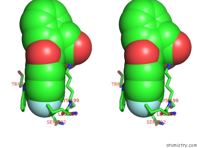
Stereo pair view
|
A full contact list of Fluorine with other atoms in the F binding
site number 1 of Human Serum Albumin in Complex with Compound 1 within 5.0Å range:
probe
|
atom
|
residue
|
distance (Å)
|
B
|
Occ
|
A:F586
b:89.5
occ:1.00
|
F11
|
A:IMX586
|
0.0
|
89.5
|
1.0
|
C6
|
A:IMX586
|
1.3
|
73.4
|
1.0
|
C4
|
A:IMX586
|
2.3
|
66.0
|
1.0
|
C7
|
A:IMX586
|
2.3
|
67.8
|
1.0
|
CD2
|
A:LEU198
|
2.9
|
48.6
|
1.0
|
CD1
|
A:LEU198
|
3.1
|
51.6
|
1.0
|
CG
|
A:LEU198
|
3.4
|
47.7
|
1.0
|
CD2
|
A:TRP214
|
3.5
|
66.0
|
1.0
|
C2
|
A:IMX586
|
3.5
|
69.3
|
1.0
|
OG
|
A:SER202
|
3.6
|
60.5
|
1.0
|
C5
|
A:IMX586
|
3.6
|
66.2
|
1.0
|
CE2
|
A:TRP214
|
3.6
|
64.0
|
1.0
|
CE3
|
A:TRP214
|
3.7
|
68.6
|
1.0
|
CB
|
A:LEU198
|
3.9
|
45.5
|
1.0
|
O
|
A:LEU198
|
3.9
|
50.1
|
1.0
|
CZ2
|
A:TRP214
|
4.0
|
69.2
|
1.0
|
CG
|
A:TRP214
|
4.0
|
55.8
|
1.0
|
CZ3
|
A:TRP214
|
4.0
|
70.5
|
1.0
|
C3
|
A:IMX586
|
4.1
|
65.8
|
1.0
|
NE1
|
A:TRP214
|
4.1
|
61.7
|
1.0
|
CH2
|
A:TRP214
|
4.1
|
72.9
|
1.0
|
C
|
A:LEU198
|
4.1
|
45.8
|
1.0
|
CD1
|
A:TRP214
|
4.3
|
56.9
|
1.0
|
N
|
A:LYS199
|
4.5
|
41.5
|
1.0
|
CG
|
A:LYS199
|
4.6
|
33.4
|
1.0
|
CA
|
A:LYS199
|
4.6
|
38.5
|
1.0
|
CA
|
A:LEU198
|
4.7
|
45.5
|
1.0
|
CB
|
A:SER202
|
4.7
|
56.3
|
1.0
|
CB
|
A:TRP214
|
4.7
|
51.0
|
1.0
|
C10
|
A:IMX586
|
4.8
|
68.0
|
1.0
|
|
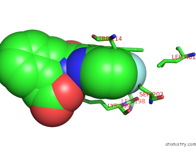
Fluorine binding site 2 out
of 4 in 3lu6
Go back to
Fluorine Binding Sites List in 3lu6
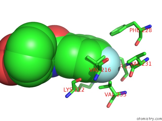
Fluorine binding site 2 out
of 4 in the Human Serum Albumin in Complex with Compound 1
Mono view
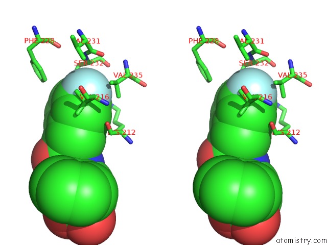
Stereo pair view
|
A full contact list of Fluorine with other atoms in the F binding
site number 2 of Human Serum Albumin in Complex with Compound 1 within 5.0Å range:
probe
|
atom
|
residue
|
distance (Å)
|
B
|
Occ
|
A:F587
b:77.0
occ:1.00
|
F11
|
A:IMX587
|
0.0
|
77.0
|
1.0
|
C6
|
A:IMX587
|
1.3
|
80.9
|
1.0
|
C4
|
A:IMX587
|
2.3
|
78.7
|
1.0
|
C7
|
A:IMX587
|
2.4
|
80.4
|
1.0
|
OG
|
A:SER232
|
3.1
|
72.8
|
1.0
|
CG2
|
A:VAL216
|
3.2
|
56.5
|
1.0
|
C2
|
A:IMX587
|
3.6
|
82.6
|
1.0
|
CA
|
A:SER232
|
3.6
|
65.0
|
1.0
|
CB
|
A:VAL216
|
3.6
|
53.9
|
1.0
|
CG1
|
A:VAL235
|
3.7
|
50.6
|
1.0
|
C5
|
A:IMX587
|
3.7
|
79.3
|
1.0
|
CB
|
A:VAL235
|
3.7
|
51.9
|
1.0
|
CG2
|
A:VAL235
|
3.8
|
51.1
|
1.0
|
CB
|
A:SER232
|
3.9
|
67.9
|
1.0
|
C3
|
A:IMX587
|
4.1
|
82.5
|
1.0
|
N
|
A:SER232
|
4.4
|
63.7
|
1.0
|
CG1
|
A:VAL216
|
4.6
|
47.6
|
1.0
|
O
|
A:SER232
|
4.6
|
65.5
|
1.0
|
C
|
A:SER232
|
4.6
|
64.8
|
1.0
|
O
|
A:LYS212
|
4.7
|
46.0
|
1.0
|
CE1
|
A:PHE228
|
4.8
|
62.4
|
1.0
|
CA
|
A:VAL216
|
4.8
|
54.0
|
1.0
|
C10
|
A:IMX587
|
4.8
|
82.2
|
1.0
|
NZ
|
A:LYS212
|
4.9
|
53.9
|
1.0
|
O
|
A:VAL231
|
4.9
|
56.7
|
1.0
|
C
|
A:VAL231
|
4.9
|
60.4
|
1.0
|
|
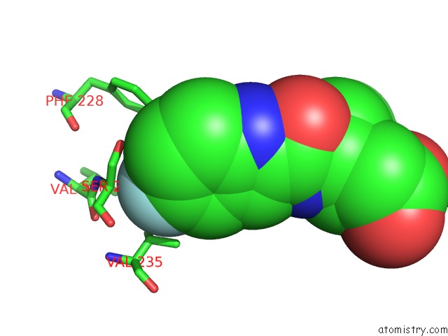
Fluorine binding site 3 out
of 4 in 3lu6
Go back to
Fluorine Binding Sites List in 3lu6
Fluorine binding site 3 out
of 4 in the Human Serum Albumin in Complex with Compound 1
Mono view
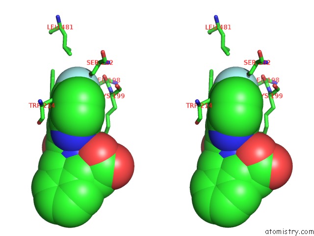
Stereo pair view
|
A full contact list of Fluorine with other atoms in the F binding
site number 3 of Human Serum Albumin in Complex with Compound 1 within 5.0Å range:
probe
|
atom
|
residue
|
distance (Å)
|
B
|
Occ
|
B:F586
b:73.7
occ:1.00
|
F11
|
B:IMX586
|
0.0
|
73.7
|
1.0
|
C6
|
B:IMX586
|
1.3
|
59.9
|
1.0
|
C7
|
B:IMX586
|
2.3
|
60.3
|
1.0
|
C4
|
B:IMX586
|
2.3
|
52.7
|
1.0
|
OG
|
B:SER202
|
2.7
|
57.3
|
1.0
|
CD2
|
B:TRP214
|
3.2
|
57.0
|
1.0
|
CG
|
B:LEU198
|
3.4
|
55.3
|
1.0
|
CE3
|
B:TRP214
|
3.5
|
65.4
|
1.0
|
CE2
|
B:TRP214
|
3.5
|
63.5
|
1.0
|
CG
|
B:TRP214
|
3.5
|
49.5
|
1.0
|
C2
|
B:IMX586
|
3.6
|
53.6
|
1.0
|
C5
|
B:IMX586
|
3.6
|
62.9
|
1.0
|
CD1
|
B:LEU198
|
3.7
|
59.5
|
1.0
|
CB
|
B:SER202
|
3.8
|
47.9
|
1.0
|
O
|
B:LEU198
|
3.9
|
49.1
|
1.0
|
NE1
|
B:TRP214
|
3.9
|
56.6
|
1.0
|
CD1
|
B:TRP214
|
3.9
|
54.4
|
1.0
|
CZ3
|
B:TRP214
|
4.0
|
65.5
|
1.0
|
CZ2
|
B:TRP214
|
4.0
|
65.2
|
1.0
|
CD2
|
B:LEU198
|
4.1
|
55.5
|
1.0
|
C3
|
B:IMX586
|
4.1
|
58.6
|
1.0
|
C
|
B:LEU198
|
4.2
|
48.6
|
1.0
|
CB
|
B:TRP214
|
4.2
|
48.2
|
1.0
|
CH2
|
B:TRP214
|
4.2
|
62.8
|
1.0
|
CB
|
B:LEU198
|
4.5
|
50.1
|
1.0
|
N
|
B:LYS199
|
4.5
|
45.9
|
1.0
|
CA
|
B:LYS199
|
4.5
|
46.2
|
1.0
|
CD2
|
B:LEU481
|
4.8
|
68.9
|
1.0
|
C10
|
B:IMX586
|
4.8
|
57.6
|
1.0
|
CA
|
B:LEU198
|
4.9
|
49.4
|
1.0
|
CG
|
B:LYS199
|
5.0
|
47.2
|
1.0
|
|
Fluorine binding site 4 out
of 4 in 3lu6
Go back to
Fluorine Binding Sites List in 3lu6
Fluorine binding site 4 out
of 4 in the Human Serum Albumin in Complex with Compound 1
Mono view
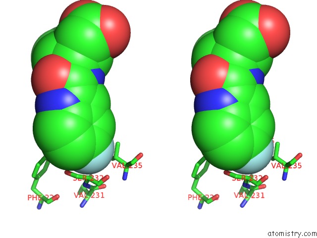
Stereo pair view
|
A full contact list of Fluorine with other atoms in the F binding
site number 4 of Human Serum Albumin in Complex with Compound 1 within 5.0Å range:
probe
|
atom
|
residue
|
distance (Å)
|
B
|
Occ
|
B:F587
b:91.0
occ:1.00
|
F11
|
B:IMX587
|
0.0
|
91.0
|
1.0
|
C6
|
B:IMX587
|
1.3
|
86.7
|
1.0
|
C4
|
B:IMX587
|
2.3
|
87.4
|
1.0
|
C7
|
B:IMX587
|
2.4
|
83.2
|
1.0
|
CG2
|
B:VAL216
|
3.1
|
53.3
|
1.0
|
CA
|
B:SER232
|
3.3
|
47.9
|
1.0
|
CG1
|
B:VAL235
|
3.4
|
44.7
|
1.0
|
CB
|
B:VAL235
|
3.5
|
46.7
|
1.0
|
CB
|
B:SER232
|
3.5
|
51.7
|
1.0
|
OG
|
B:SER232
|
3.6
|
61.3
|
1.0
|
C2
|
B:IMX587
|
3.6
|
85.7
|
1.0
|
C5
|
B:IMX587
|
3.6
|
86.7
|
1.0
|
CG2
|
B:VAL235
|
3.9
|
47.4
|
1.0
|
C3
|
B:IMX587
|
4.1
|
86.9
|
1.0
|
O
|
B:SER232
|
4.1
|
48.2
|
1.0
|
N
|
B:SER232
|
4.2
|
47.2
|
1.0
|
C
|
B:SER232
|
4.2
|
49.2
|
1.0
|
CB
|
B:VAL216
|
4.4
|
45.7
|
1.0
|
CG1
|
B:VAL216
|
4.6
|
51.3
|
1.0
|
CZ
|
B:PHE228
|
4.8
|
45.9
|
1.0
|
CA
|
B:VAL235
|
4.8
|
43.8
|
1.0
|
C10
|
B:IMX587
|
4.8
|
83.3
|
1.0
|
CE1
|
B:PHE228
|
4.9
|
46.8
|
1.0
|
C
|
B:VAL231
|
4.9
|
48.1
|
1.0
|
O
|
B:VAL231
|
4.9
|
47.7
|
1.0
|
|
Reference:
D.Buttar,
N.Colclough,
S.Gerhardt,
P.A.Macfaul,
S.D.Phillips,
A.Plowright,
P.Whittamore,
K.Tam,
K.Maskos,
S.Steinbacher,
H.Steuber.
A Combined Spectroscopic and Crystallographic Approach to Probing Drug-Human Serum Albumin Interactions Bioorg.Med.Chem. V. 18 7486 2010.
ISSN: ISSN 0968-0896
PubMed: 20869876
DOI: 10.1016/J.BMC.2010.08.052
Page generated: Wed Jul 31 20:35:37 2024
|