Fluorine »
PDB 3lz3-3n0n »
3m2w »
Fluorine in PDB 3m2w: Crystal Structure of Mapkak Kinase 2 (MK2) Complexed with A Spiroazetidine-Tetracyclic Atp Site Inhibitor
Enzymatic activity of Crystal Structure of Mapkak Kinase 2 (MK2) Complexed with A Spiroazetidine-Tetracyclic Atp Site Inhibitor
All present enzymatic activity of Crystal Structure of Mapkak Kinase 2 (MK2) Complexed with A Spiroazetidine-Tetracyclic Atp Site Inhibitor:
2.7.11.1;
2.7.11.1;
Protein crystallography data
The structure of Crystal Structure of Mapkak Kinase 2 (MK2) Complexed with A Spiroazetidine-Tetracyclic Atp Site Inhibitor, PDB code: 3m2w
was solved by
M.Kroemer,
L.Revesz,
C.Be,
A.Izaac,
C.Huppertz,
A.Schlapbach,
C.Scheufler,
with X-Ray Crystallography technique. A brief refinement statistics is given in the table below:
| Resolution Low / High (Å) | 29.26 / 2.41 |
| Space group | P 63 2 2 |
| Cell size a, b, c (Å), α, β, γ (°) | 102.982, 102.982, 165.433, 90.00, 90.00, 120.00 |
| R / Rfree (%) | 18.5 / 22.7 |
Other elements in 3m2w:
The structure of Crystal Structure of Mapkak Kinase 2 (MK2) Complexed with A Spiroazetidine-Tetracyclic Atp Site Inhibitor also contains other interesting chemical elements:
| Magnesium | (Mg) | 1 atom |
Fluorine Binding Sites:
The binding sites of Fluorine atom in the Crystal Structure of Mapkak Kinase 2 (MK2) Complexed with A Spiroazetidine-Tetracyclic Atp Site Inhibitor
(pdb code 3m2w). This binding sites where shown within
5.0 Angstroms radius around Fluorine atom.
In total only one binding site of Fluorine was determined in the Crystal Structure of Mapkak Kinase 2 (MK2) Complexed with A Spiroazetidine-Tetracyclic Atp Site Inhibitor, PDB code: 3m2w:
In total only one binding site of Fluorine was determined in the Crystal Structure of Mapkak Kinase 2 (MK2) Complexed with A Spiroazetidine-Tetracyclic Atp Site Inhibitor, PDB code: 3m2w:
Fluorine binding site 1 out of 1 in 3m2w
Go back to
Fluorine binding site 1 out
of 1 in the Crystal Structure of Mapkak Kinase 2 (MK2) Complexed with A Spiroazetidine-Tetracyclic Atp Site Inhibitor

Mono view
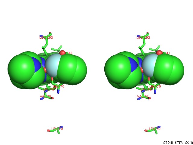
Stereo pair view
Mono view
Stereo pair view
A full contact list of Fluorine with other atoms in the F binding
site number 1 of Crystal Structure of Mapkak Kinase 2 (MK2) Complexed with A Spiroazetidine-Tetracyclic Atp Site Inhibitor within 5.0Å range:
|
Reference:
L.Revesz,
A.Schlapbach,
R.Aichholz,
J.Dawson,
R.Feifel,
S.Hawtin,
A.Littlewood-Evans,
G.Koch,
M.Kroemer,
H.Mobitz,
C.Scheufler,
J.Velcicky,
C.Huppertz.
In Vivo and in Vitro Sar of Tetracyclic Mapkap-K2 (MK2) Inhibitors. Part II. Bioorg.Med.Chem.Lett. V. 20 4719 2010.
ISSN: ISSN 0960-894X
PubMed: 20591669
DOI: 10.1016/J.BMCL.2010.04.023
Page generated: Wed Jul 31 20:39:03 2024
ISSN: ISSN 0960-894X
PubMed: 20591669
DOI: 10.1016/J.BMCL.2010.04.023
Last articles
Zn in 9J0NZn in 9J0O
Zn in 9J0P
Zn in 9FJX
Zn in 9EKB
Zn in 9C0F
Zn in 9CAH
Zn in 9CH0
Zn in 9CH3
Zn in 9CH1