Fluorine »
PDB 3lz3-3n0n »
3m4h »
Fluorine in PDB 3m4h: Human Aldose Reductase Mutant T113V Complexed with IDD388
Enzymatic activity of Human Aldose Reductase Mutant T113V Complexed with IDD388
All present enzymatic activity of Human Aldose Reductase Mutant T113V Complexed with IDD388:
1.1.1.21;
1.1.1.21;
Protein crystallography data
The structure of Human Aldose Reductase Mutant T113V Complexed with IDD388, PDB code: 3m4h
was solved by
C.Koch,
A.Heine,
G.Klebe,
with X-Ray Crystallography technique. A brief refinement statistics is given in the table below:
| Resolution Low / High (Å) | 10.00 / 0.94 |
| Space group | P 1 21 1 |
| Cell size a, b, c (Å), α, β, γ (°) | 49.390, 66.880, 47.370, 90.00, 92.11, 90.00 |
| R / Rfree (%) | 10.3 / 12.2 |
Other elements in 3m4h:
The structure of Human Aldose Reductase Mutant T113V Complexed with IDD388 also contains other interesting chemical elements:
| Bromine | (Br) | 2 atoms |
| Chlorine | (Cl) | 1 atom |
Fluorine Binding Sites:
The binding sites of Fluorine atom in the Human Aldose Reductase Mutant T113V Complexed with IDD388
(pdb code 3m4h). This binding sites where shown within
5.0 Angstroms radius around Fluorine atom.
In total only one binding site of Fluorine was determined in the Human Aldose Reductase Mutant T113V Complexed with IDD388, PDB code: 3m4h:
In total only one binding site of Fluorine was determined in the Human Aldose Reductase Mutant T113V Complexed with IDD388, PDB code: 3m4h:
Fluorine binding site 1 out of 1 in 3m4h
Go back to
Fluorine binding site 1 out
of 1 in the Human Aldose Reductase Mutant T113V Complexed with IDD388
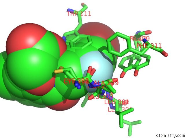
Mono view
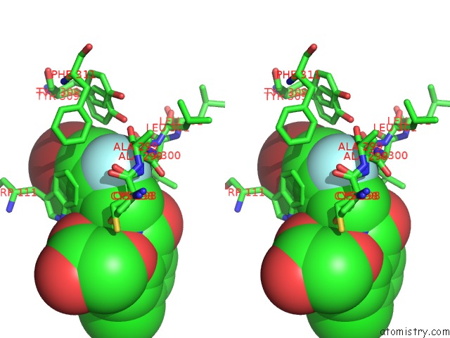
Stereo pair view
Mono view
Stereo pair view
A full contact list of Fluorine with other atoms in the F binding
site number 1 of Human Aldose Reductase Mutant T113V Complexed with IDD388 within 5.0Å range:
|
Reference:
C.Koch,
A.Heine,
G.Klebe.
Tracing the Detail: How Mutations Affect Binding Modes and Thermodynamic Signatures of Closely Related Aldose Reductase Inhibitors J.Mol.Biol. V. 406 700 2011.
ISSN: ISSN 0022-2836
PubMed: 21185307
DOI: 10.1016/J.JMB.2010.11.058
Page generated: Wed Jul 31 20:39:46 2024
ISSN: ISSN 0022-2836
PubMed: 21185307
DOI: 10.1016/J.JMB.2010.11.058
Last articles
Zn in 9J0NZn in 9J0O
Zn in 9J0P
Zn in 9FJX
Zn in 9EKB
Zn in 9C0F
Zn in 9CAH
Zn in 9CH0
Zn in 9CH3
Zn in 9CH1