Fluorine »
PDB 3lz3-3n0n »
3mhz »
Fluorine in PDB 3mhz: 1.7A Structure of 2-Fluorohistidine Labeled Protective Antigen
Protein crystallography data
The structure of 1.7A Structure of 2-Fluorohistidine Labeled Protective Antigen, PDB code: 3mhz
was solved by
S.Lovell,
K.P.Battaile,
D.S.Wimalasena,
B.E.Janowiak,
M.Miyagi,
J.Sun,
J.Hajduch,
C.Pooput,
K.L.Kirk,
J.G.Bann,
with X-Ray Crystallography technique. A brief refinement statistics is given in the table below:
| Resolution Low / High (Å) | 50.00 / 1.70 |
| Space group | P 21 21 21 |
| Cell size a, b, c (Å), α, β, γ (°) | 71.368, 93.975, 119.115, 90.00, 90.00, 90.00 |
| R / Rfree (%) | 19.2 / 22.2 |
Other elements in 3mhz:
The structure of 1.7A Structure of 2-Fluorohistidine Labeled Protective Antigen also contains other interesting chemical elements:
| Calcium | (Ca) | 2 atoms |
Fluorine Binding Sites:
The binding sites of Fluorine atom in the 1.7A Structure of 2-Fluorohistidine Labeled Protective Antigen
(pdb code 3mhz). This binding sites where shown within
5.0 Angstroms radius around Fluorine atom.
In total 7 binding sites of Fluorine where determined in the 1.7A Structure of 2-Fluorohistidine Labeled Protective Antigen, PDB code: 3mhz:
Jump to Fluorine binding site number: 1; 2; 3; 4; 5; 6; 7;
In total 7 binding sites of Fluorine where determined in the 1.7A Structure of 2-Fluorohistidine Labeled Protective Antigen, PDB code: 3mhz:
Jump to Fluorine binding site number: 1; 2; 3; 4; 5; 6; 7;
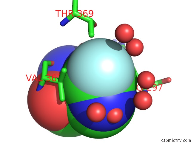
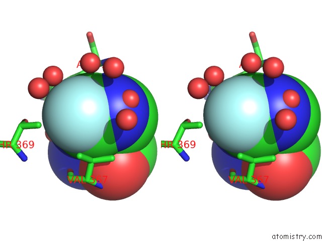
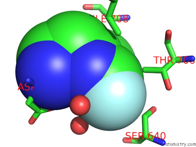
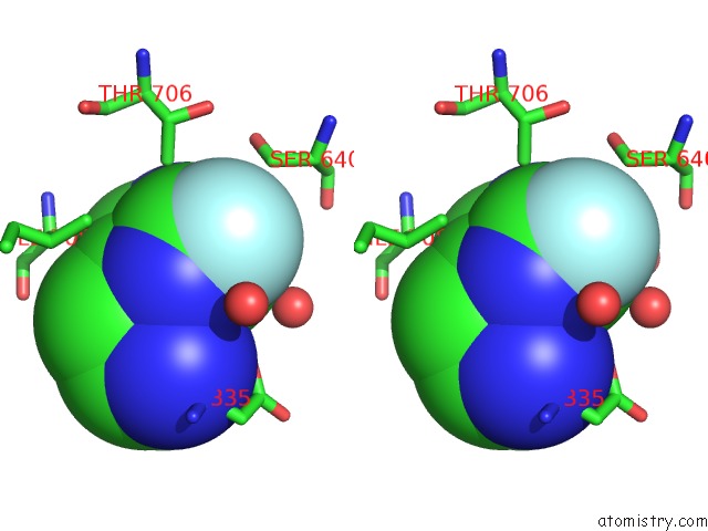
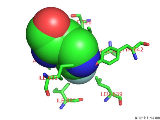
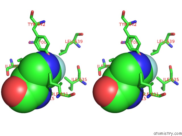
Fluorine binding site 1 out of 7 in 3mhz
Go back to
Fluorine binding site 1 out
of 7 in the 1.7A Structure of 2-Fluorohistidine Labeled Protective Antigen
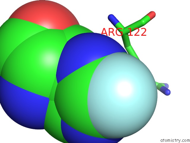
Mono view
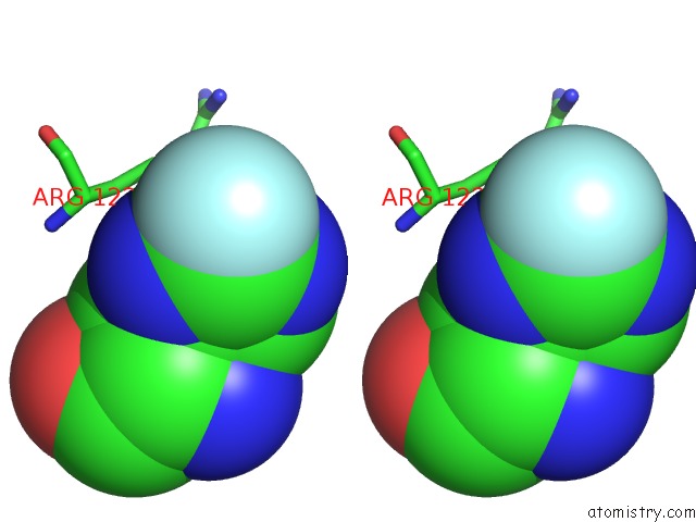
Stereo pair view
Mono view
Stereo pair view
A full contact list of Fluorine with other atoms in the F binding
site number 1 of 1.7A Structure of 2-Fluorohistidine Labeled Protective Antigen within 5.0Å range:
|
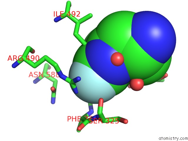
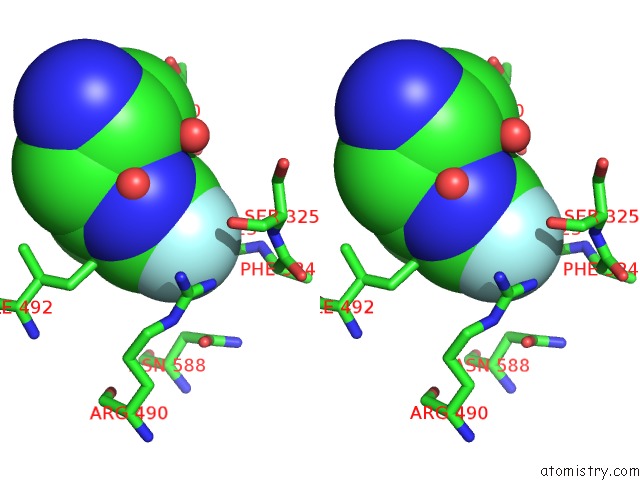
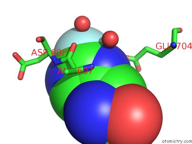
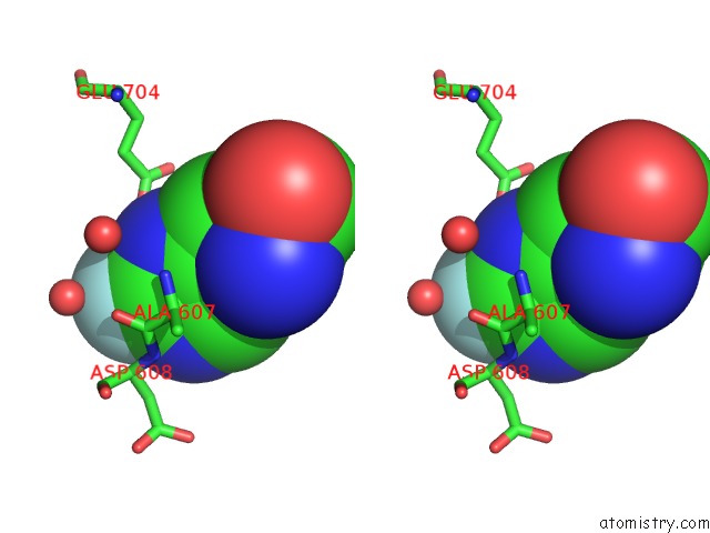
Fluorine binding site 2 out of 7 in 3mhz
Go back to
Fluorine binding site 2 out
of 7 in the 1.7A Structure of 2-Fluorohistidine Labeled Protective Antigen
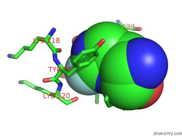
Mono view
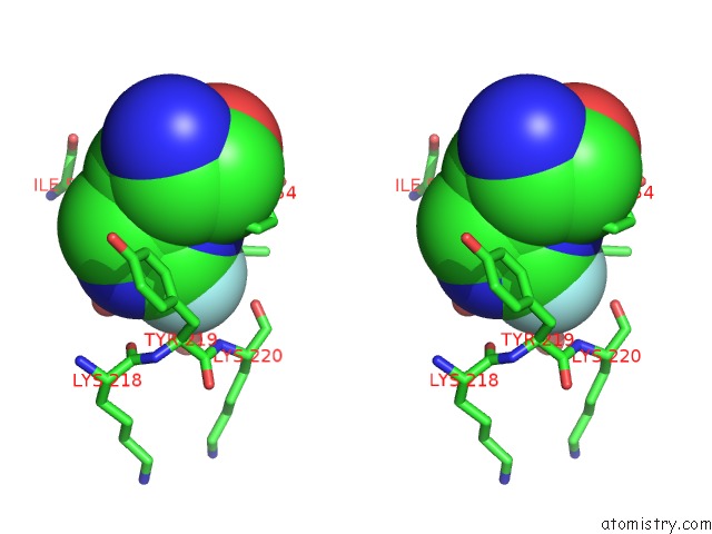
Stereo pair view
Mono view
Stereo pair view
A full contact list of Fluorine with other atoms in the F binding
site number 2 of 1.7A Structure of 2-Fluorohistidine Labeled Protective Antigen within 5.0Å range:
|
Fluorine binding site 3 out of 7 in 3mhz
Go back to
Fluorine binding site 3 out
of 7 in the 1.7A Structure of 2-Fluorohistidine Labeled Protective Antigen
Mono view
Stereo pair view
Mono view
Stereo pair view
A full contact list of Fluorine with other atoms in the F binding
site number 3 of 1.7A Structure of 2-Fluorohistidine Labeled Protective Antigen within 5.0Å range:
|
Fluorine binding site 4 out of 7 in 3mhz
Go back to
Fluorine binding site 4 out
of 7 in the 1.7A Structure of 2-Fluorohistidine Labeled Protective Antigen
Mono view
Stereo pair view
Mono view
Stereo pair view
A full contact list of Fluorine with other atoms in the F binding
site number 4 of 1.7A Structure of 2-Fluorohistidine Labeled Protective Antigen within 5.0Å range:
|
Fluorine binding site 5 out of 7 in 3mhz
Go back to
Fluorine binding site 5 out
of 7 in the 1.7A Structure of 2-Fluorohistidine Labeled Protective Antigen
Mono view
Stereo pair view
Mono view
Stereo pair view
A full contact list of Fluorine with other atoms in the F binding
site number 5 of 1.7A Structure of 2-Fluorohistidine Labeled Protective Antigen within 5.0Å range:
|
Fluorine binding site 6 out of 7 in 3mhz
Go back to
Fluorine binding site 6 out
of 7 in the 1.7A Structure of 2-Fluorohistidine Labeled Protective Antigen
Mono view
Stereo pair view
Mono view
Stereo pair view
A full contact list of Fluorine with other atoms in the F binding
site number 6 of 1.7A Structure of 2-Fluorohistidine Labeled Protective Antigen within 5.0Å range:
|
Fluorine binding site 7 out of 7 in 3mhz
Go back to
Fluorine binding site 7 out
of 7 in the 1.7A Structure of 2-Fluorohistidine Labeled Protective Antigen
Mono view
Stereo pair view
Mono view
Stereo pair view
A full contact list of Fluorine with other atoms in the F binding
site number 7 of 1.7A Structure of 2-Fluorohistidine Labeled Protective Antigen within 5.0Å range:
|
Reference:
D.S.Wimalasena,
B.E.Janowiak,
S.Lovell,
M.Miyagi,
J.Sun,
H.Zhou,
J.Hajduch,
C.Pooput,
K.L.Kirk,
K.P.Battaile,
J.G.Bann.
Evidence That Histidine Protonation of Receptor-Bound Anthrax Protective Antigen Is A Trigger For Pore Formation. Biochemistry V. 49 6973 2010.
ISSN: ISSN 0006-2960
PubMed: 20672855
DOI: 10.1021/BI100647Z
Page generated: Mon Jul 14 17:54:33 2025
ISSN: ISSN 0006-2960
PubMed: 20672855
DOI: 10.1021/BI100647Z
Last articles
Fe in 2YXOFe in 2YRS
Fe in 2YXC
Fe in 2YNM
Fe in 2YVJ
Fe in 2YP1
Fe in 2YU2
Fe in 2YU1
Fe in 2YQB
Fe in 2YOO