Fluorine »
PDB 3lz3-3n0n »
3mne »
Fluorine in PDB 3mne: Crystal Structure of the Agonist Form of Mouse Glucocorticoid Receptor Stabilized By F608S Mutation at 1.96A
Protein crystallography data
The structure of Crystal Structure of the Agonist Form of Mouse Glucocorticoid Receptor Stabilized By F608S Mutation at 1.96A, PDB code: 3mne
was solved by
G.A.Schoch,
T.Seitz,
J.Benz,
D.Banner,
M.Stihle,
B.D'arcy,
R.Thoma,
R.Sterner,
M.Hennig,
A.Ruf,
with X-Ray Crystallography technique. A brief refinement statistics is given in the table below:
| Resolution Low / High (Å) | 12.98 / 1.96 |
| Space group | P 65 |
| Cell size a, b, c (Å), α, β, γ (°) | 71.820, 71.820, 128.650, 90.00, 90.00, 120.00 |
| R / Rfree (%) | 15.1 / 18.5 |
Fluorine Binding Sites:
The binding sites of Fluorine atom in the Crystal Structure of the Agonist Form of Mouse Glucocorticoid Receptor Stabilized By F608S Mutation at 1.96A
(pdb code 3mne). This binding sites where shown within
5.0 Angstroms radius around Fluorine atom.
In total only one binding site of Fluorine was determined in the Crystal Structure of the Agonist Form of Mouse Glucocorticoid Receptor Stabilized By F608S Mutation at 1.96A, PDB code: 3mne:
In total only one binding site of Fluorine was determined in the Crystal Structure of the Agonist Form of Mouse Glucocorticoid Receptor Stabilized By F608S Mutation at 1.96A, PDB code: 3mne:
Fluorine binding site 1 out of 1 in 3mne
Go back to
Fluorine binding site 1 out
of 1 in the Crystal Structure of the Agonist Form of Mouse Glucocorticoid Receptor Stabilized By F608S Mutation at 1.96A

Mono view
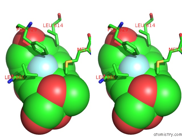
Stereo pair view
Mono view
Stereo pair view
A full contact list of Fluorine with other atoms in the F binding
site number 1 of Crystal Structure of the Agonist Form of Mouse Glucocorticoid Receptor Stabilized By F608S Mutation at 1.96A within 5.0Å range:
|
Reference:
T.Seitz,
R.Thoma,
G.A.Schoch,
M.Stihle,
J.Benz,
B.D'arcy,
A.Wiget,
A.Ruf,
M.Hennig,
R.Sterner.
Enhancing the Stability and Solubility of the Glucocorticoid Receptor Ligand-Binding Domain By High-Throughput Library Screening. J.Mol.Biol. V. 403 562 2010.
ISSN: ISSN 0022-2836
PubMed: 20850457
DOI: 10.1016/J.JMB.2010.08.048
Page generated: Wed Jul 31 20:42:56 2024
ISSN: ISSN 0022-2836
PubMed: 20850457
DOI: 10.1016/J.JMB.2010.08.048
Last articles
Zn in 9J0NZn in 9J0O
Zn in 9J0P
Zn in 9FJX
Zn in 9EKB
Zn in 9C0F
Zn in 9CAH
Zn in 9CH0
Zn in 9CH3
Zn in 9CH1