Fluorine »
PDB 3lz3-3n0n »
3mxe »
Fluorine in PDB 3mxe: Crystal Structure of Hiv-1 Protease Inhibitor, KC32 Complexed with Wild-Type Protease
Enzymatic activity of Crystal Structure of Hiv-1 Protease Inhibitor, KC32 Complexed with Wild-Type Protease
All present enzymatic activity of Crystal Structure of Hiv-1 Protease Inhibitor, KC32 Complexed with Wild-Type Protease:
3.4.23.16;
3.4.23.16;
Protein crystallography data
The structure of Crystal Structure of Hiv-1 Protease Inhibitor, KC32 Complexed with Wild-Type Protease, PDB code: 3mxe
was solved by
M.N.L.Nalam,
C.A.Schiffer,
with X-Ray Crystallography technique. A brief refinement statistics is given in the table below:
| Resolution Low / High (Å) | 39.28 / 1.85 |
| Space group | P 21 21 21 |
| Cell size a, b, c (Å), α, β, γ (°) | 50.790, 58.245, 62.014, 90.00, 90.00, 90.00 |
| R / Rfree (%) | 18 / 22.3 |
Fluorine Binding Sites:
The binding sites of Fluorine atom in the Crystal Structure of Hiv-1 Protease Inhibitor, KC32 Complexed with Wild-Type Protease
(pdb code 3mxe). This binding sites where shown within
5.0 Angstroms radius around Fluorine atom.
In total 3 binding sites of Fluorine where determined in the Crystal Structure of Hiv-1 Protease Inhibitor, KC32 Complexed with Wild-Type Protease, PDB code: 3mxe:
Jump to Fluorine binding site number: 1; 2; 3;
In total 3 binding sites of Fluorine where determined in the Crystal Structure of Hiv-1 Protease Inhibitor, KC32 Complexed with Wild-Type Protease, PDB code: 3mxe:
Jump to Fluorine binding site number: 1; 2; 3;
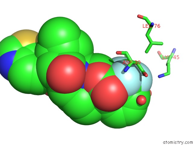
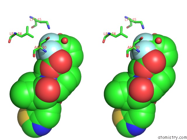
Fluorine binding site 1 out of 3 in 3mxe
Go back to
Fluorine binding site 1 out
of 3 in the Crystal Structure of Hiv-1 Protease Inhibitor, KC32 Complexed with Wild-Type Protease
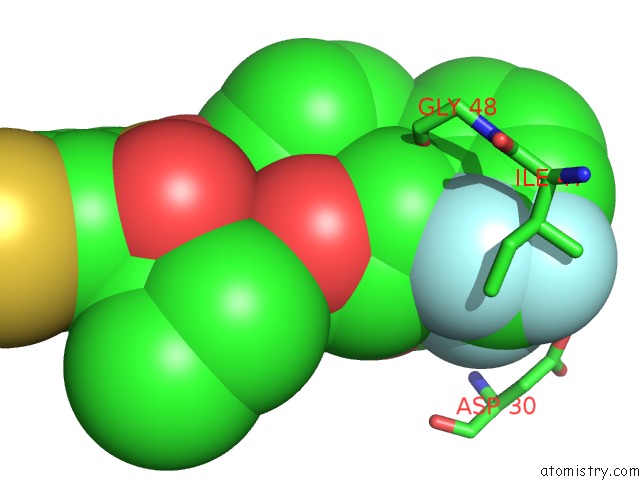
Mono view
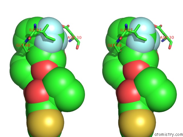
Stereo pair view
Mono view
Stereo pair view
A full contact list of Fluorine with other atoms in the F binding
site number 1 of Crystal Structure of Hiv-1 Protease Inhibitor, KC32 Complexed with Wild-Type Protease within 5.0Å range:
|
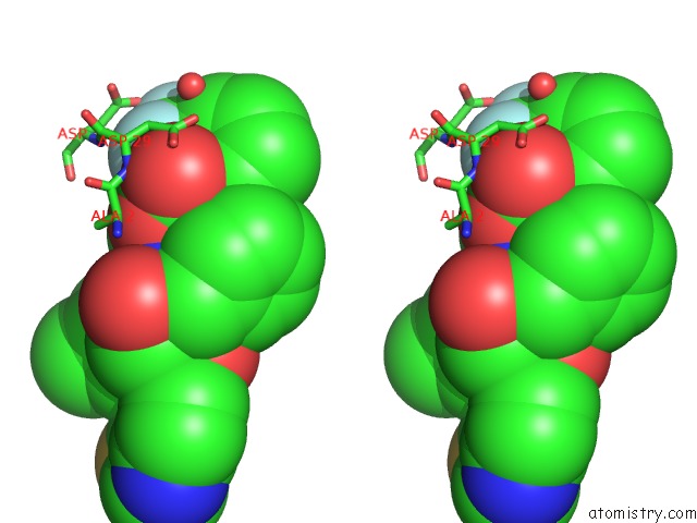
Fluorine binding site 2 out of 3 in 3mxe
Go back to
Fluorine binding site 2 out
of 3 in the Crystal Structure of Hiv-1 Protease Inhibitor, KC32 Complexed with Wild-Type Protease
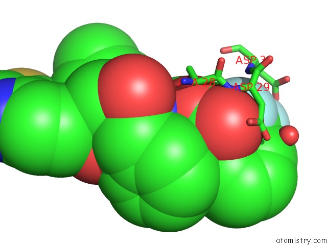
Mono view
Stereo pair view
Mono view
Stereo pair view
A full contact list of Fluorine with other atoms in the F binding
site number 2 of Crystal Structure of Hiv-1 Protease Inhibitor, KC32 Complexed with Wild-Type Protease within 5.0Å range:
|
Fluorine binding site 3 out of 3 in 3mxe
Go back to
Fluorine binding site 3 out
of 3 in the Crystal Structure of Hiv-1 Protease Inhibitor, KC32 Complexed with Wild-Type Protease
Mono view
Stereo pair view
Mono view
Stereo pair view
A full contact list of Fluorine with other atoms in the F binding
site number 3 of Crystal Structure of Hiv-1 Protease Inhibitor, KC32 Complexed with Wild-Type Protease within 5.0Å range:
|
Reference:
A.Ali,
G.S.Reddy,
M.N.Nalam,
S.G.Anjum,
H.Cao,
C.A.Schiffer,
T.M.Rana.
Structure-Based Design, Synthesis, and Structure-Activity Relationship Studies of Hiv-1 Protease Inhibitors Incorporating Phenyloxazolidinones. J.Med.Chem. V. 53 7699 2010.
ISSN: ISSN 0022-2623
PubMed: 20958050
DOI: 10.1021/JM1008743
Page generated: Mon Jul 14 17:57:44 2025
ISSN: ISSN 0022-2623
PubMed: 20958050
DOI: 10.1021/JM1008743
Last articles
Fe in 2YXOFe in 2YRS
Fe in 2YXC
Fe in 2YNM
Fe in 2YVJ
Fe in 2YP1
Fe in 2YU2
Fe in 2YU1
Fe in 2YQB
Fe in 2YOO