Fluorine »
PDB 3n2m-3nly »
3n4b »
Fluorine in PDB 3n4b: Crystal Structure of Human Carbonic Anhydrase II in Complex with A Benzenesulfonamide Inhibitor
Enzymatic activity of Crystal Structure of Human Carbonic Anhydrase II in Complex with A Benzenesulfonamide Inhibitor
All present enzymatic activity of Crystal Structure of Human Carbonic Anhydrase II in Complex with A Benzenesulfonamide Inhibitor:
4.2.1.1;
4.2.1.1;
Protein crystallography data
The structure of Crystal Structure of Human Carbonic Anhydrase II in Complex with A Benzenesulfonamide Inhibitor, PDB code: 3n4b
was solved by
B.S.Avvaru,
J.Wagner,
A.H.Robbins,
R.Mckenna,
with X-Ray Crystallography technique. A brief refinement statistics is given in the table below:
| Resolution Low / High (Å) | 19.85 / 1.60 |
| Space group | P 1 21 1 |
| Cell size a, b, c (Å), α, β, γ (°) | 42.465, 41.414, 72.037, 90.00, 104.13, 90.00 |
| R / Rfree (%) | 14.1 / 17 |
Other elements in 3n4b:
The structure of Crystal Structure of Human Carbonic Anhydrase II in Complex with A Benzenesulfonamide Inhibitor also contains other interesting chemical elements:
| Zinc | (Zn) | 1 atom |
Fluorine Binding Sites:
The binding sites of Fluorine atom in the Crystal Structure of Human Carbonic Anhydrase II in Complex with A Benzenesulfonamide Inhibitor
(pdb code 3n4b). This binding sites where shown within
5.0 Angstroms radius around Fluorine atom.
In total only one binding site of Fluorine was determined in the Crystal Structure of Human Carbonic Anhydrase II in Complex with A Benzenesulfonamide Inhibitor, PDB code: 3n4b:
In total only one binding site of Fluorine was determined in the Crystal Structure of Human Carbonic Anhydrase II in Complex with A Benzenesulfonamide Inhibitor, PDB code: 3n4b:
Fluorine binding site 1 out of 1 in 3n4b
Go back to
Fluorine binding site 1 out
of 1 in the Crystal Structure of Human Carbonic Anhydrase II in Complex with A Benzenesulfonamide Inhibitor
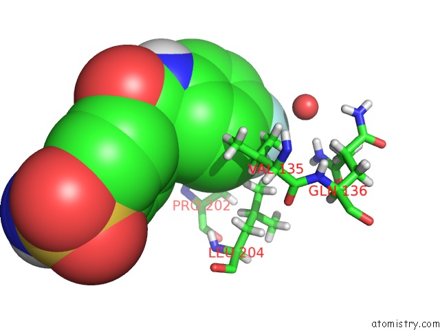
Mono view
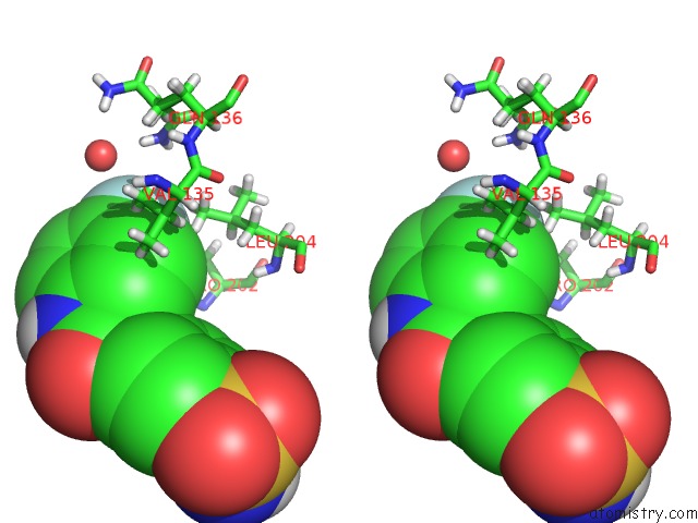
Stereo pair view
Mono view
Stereo pair view
A full contact list of Fluorine with other atoms in the F binding
site number 1 of Crystal Structure of Human Carbonic Anhydrase II in Complex with A Benzenesulfonamide Inhibitor within 5.0Å range:
|
Reference:
F.Pacchiano,
M.Aggarwal,
B.S.Avvaru,
A.H.Robbins,
A.Scozzafava,
R.Mckenna,
C.T.Supuran.
Selective Hydrophobic Pocket Binding Observed Within the Carbonic Anhydrase II Active Site Accommodate Different 4-Substituted-Ureido-Benzenesulfonamides and Correlate to Inhibitor Potency. Chem.Commun.(Camb.) V. 46 8371 2010.
ISSN: ISSN 1359-7345
PubMed: 20922253
DOI: 10.1039/C0CC02707C
Page generated: Wed Jul 31 20:50:01 2024
ISSN: ISSN 1359-7345
PubMed: 20922253
DOI: 10.1039/C0CC02707C
Last articles
Cl in 5RSLCl in 5ROW
Cl in 5RMJ
Cl in 5ROP
Cl in 5RML
Cl in 5RKR
Cl in 5RKE
Cl in 5RH4
Cl in 5RH3
Cl in 5RH2