Fluorine »
PDB 3n2m-3nly »
3n8w »
Fluorine in PDB 3n8w: Crystal Structure of R120Q/Native Cyclooxygenase-1 Heterodimer Mutant in Complex with Flurbiprofen
Enzymatic activity of Crystal Structure of R120Q/Native Cyclooxygenase-1 Heterodimer Mutant in Complex with Flurbiprofen
All present enzymatic activity of Crystal Structure of R120Q/Native Cyclooxygenase-1 Heterodimer Mutant in Complex with Flurbiprofen:
1.14.99.1;
1.14.99.1;
Protein crystallography data
The structure of Crystal Structure of R120Q/Native Cyclooxygenase-1 Heterodimer Mutant in Complex with Flurbiprofen, PDB code: 3n8w
was solved by
R.S.Sidhu,
with X-Ray Crystallography technique. A brief refinement statistics is given in the table below:
| Resolution Low / High (Å) | 46.91 / 2.75 |
| Space group | P 65 |
| Cell size a, b, c (Å), α, β, γ (°) | 182.483, 182.483, 103.095, 90.00, 90.00, 120.00 |
| R / Rfree (%) | 17.8 / 20.1 |
Other elements in 3n8w:
The structure of Crystal Structure of R120Q/Native Cyclooxygenase-1 Heterodimer Mutant in Complex with Flurbiprofen also contains other interesting chemical elements:
| Iron | (Fe) | 2 atoms |
Fluorine Binding Sites:
The binding sites of Fluorine atom in the Crystal Structure of R120Q/Native Cyclooxygenase-1 Heterodimer Mutant in Complex with Flurbiprofen
(pdb code 3n8w). This binding sites where shown within
5.0 Angstroms radius around Fluorine atom.
In total only one binding site of Fluorine was determined in the Crystal Structure of R120Q/Native Cyclooxygenase-1 Heterodimer Mutant in Complex with Flurbiprofen, PDB code: 3n8w:
In total only one binding site of Fluorine was determined in the Crystal Structure of R120Q/Native Cyclooxygenase-1 Heterodimer Mutant in Complex with Flurbiprofen, PDB code: 3n8w:
Fluorine binding site 1 out of 1 in 3n8w
Go back to
Fluorine binding site 1 out
of 1 in the Crystal Structure of R120Q/Native Cyclooxygenase-1 Heterodimer Mutant in Complex with Flurbiprofen
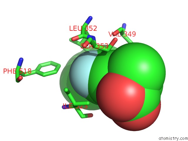
Mono view
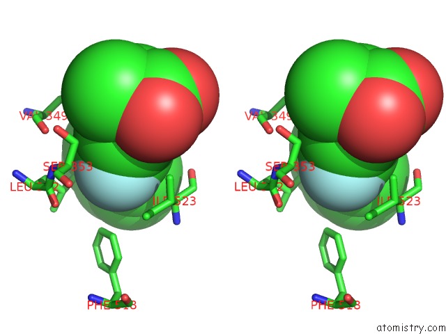
Stereo pair view
Mono view
Stereo pair view
A full contact list of Fluorine with other atoms in the F binding
site number 1 of Crystal Structure of R120Q/Native Cyclooxygenase-1 Heterodimer Mutant in Complex with Flurbiprofen within 5.0Å range:
|
Reference:
R.S.Sidhu,
J.Y.Lee,
C.Yuan,
W.L.Smith.
Comparison of Cyclooxygenase-1 Crystal Structures: Cross-Talk Between Monomers Comprising Cyclooxygenase-1 Homodimers Biochemistry V. 49 7069 2010.
ISSN: ISSN 0006-2960
PubMed: 20669977
DOI: 10.1021/BI1003298
Page generated: Wed Jul 31 20:51:02 2024
ISSN: ISSN 0006-2960
PubMed: 20669977
DOI: 10.1021/BI1003298
Last articles
Zn in 9MJ5Zn in 9HNW
Zn in 9G0L
Zn in 9FNE
Zn in 9DZN
Zn in 9E0I
Zn in 9D32
Zn in 9DAK
Zn in 8ZXC
Zn in 8ZUF