Fluorine »
PDB 3n2m-3nly »
3nk8 »
Fluorine in PDB 3nk8: Trypsin in Complex with Fluorine-Containing Fragment
Enzymatic activity of Trypsin in Complex with Fluorine-Containing Fragment
All present enzymatic activity of Trypsin in Complex with Fluorine-Containing Fragment:
3.4.21.4;
3.4.21.4;
Protein crystallography data
The structure of Trypsin in Complex with Fluorine-Containing Fragment, PDB code: 3nk8
was solved by
N.Schiering,
A.Vulpetti,
C.Dalvit,
with X-Ray Crystallography technique. A brief refinement statistics is given in the table below:
| Resolution Low / High (Å) | 33.30 / 1.15 |
| Space group | P 21 21 21 |
| Cell size a, b, c (Å), α, β, γ (°) | 54.720, 58.266, 66.770, 90.00, 90.00, 90.00 |
| R / Rfree (%) | 12.2 / 14.3 |
Other elements in 3nk8:
The structure of Trypsin in Complex with Fluorine-Containing Fragment also contains other interesting chemical elements:
| Calcium | (Ca) | 1 atom |
Fluorine Binding Sites:
The binding sites of Fluorine atom in the Trypsin in Complex with Fluorine-Containing Fragment
(pdb code 3nk8). This binding sites where shown within
5.0 Angstroms radius around Fluorine atom.
In total 3 binding sites of Fluorine where determined in the Trypsin in Complex with Fluorine-Containing Fragment, PDB code: 3nk8:
Jump to Fluorine binding site number: 1; 2; 3;
In total 3 binding sites of Fluorine where determined in the Trypsin in Complex with Fluorine-Containing Fragment, PDB code: 3nk8:
Jump to Fluorine binding site number: 1; 2; 3;
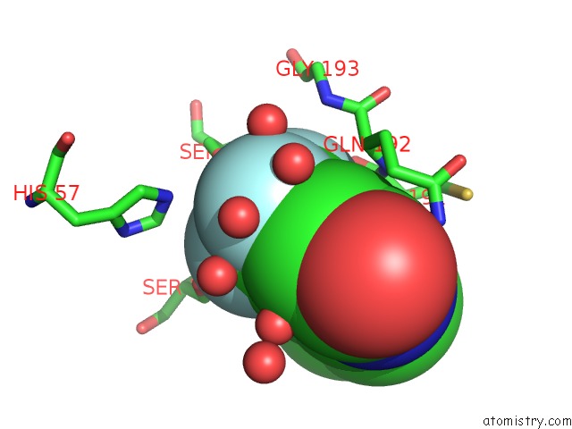
Fluorine binding site 1 out of 3 in 3nk8
Go back to
Fluorine binding site 1 out
of 3 in the Trypsin in Complex with Fluorine-Containing Fragment
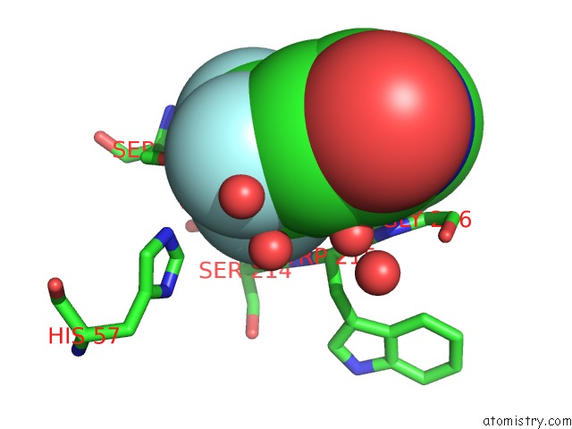
Mono view
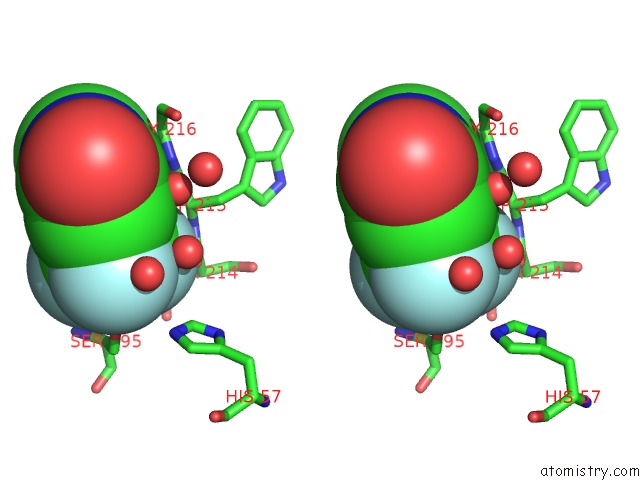
Stereo pair view
Mono view
Stereo pair view
A full contact list of Fluorine with other atoms in the F binding
site number 1 of Trypsin in Complex with Fluorine-Containing Fragment within 5.0Å range:
|
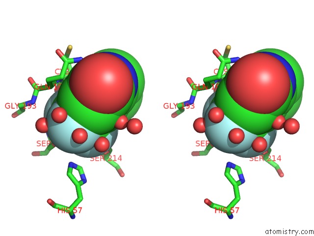
Fluorine binding site 2 out of 3 in 3nk8
Go back to
Fluorine binding site 2 out
of 3 in the Trypsin in Complex with Fluorine-Containing Fragment
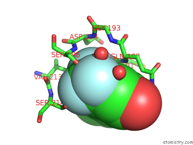
Mono view
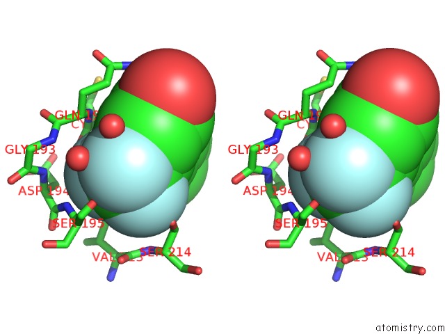
Stereo pair view
Mono view
Stereo pair view
A full contact list of Fluorine with other atoms in the F binding
site number 2 of Trypsin in Complex with Fluorine-Containing Fragment within 5.0Å range:
|
Fluorine binding site 3 out of 3 in 3nk8
Go back to
Fluorine binding site 3 out
of 3 in the Trypsin in Complex with Fluorine-Containing Fragment
Mono view
Stereo pair view
Mono view
Stereo pair view
A full contact list of Fluorine with other atoms in the F binding
site number 3 of Trypsin in Complex with Fluorine-Containing Fragment within 5.0Å range:
|
Reference:
A.Vulpetti,
N.Schiering,
C.Dalvit.
Combined Use of Computational Chemistry, uc(Nmr) Screening, and X-Ray Crystallography For Identification and Characterization of Fluorophilic Protein Environments. Proteins V. 78 3281 2010.
ISSN: ISSN 0887-3585
PubMed: 20886466
DOI: 10.1002/PROT.22836
Page generated: Wed Jul 31 20:53:23 2024
ISSN: ISSN 0887-3585
PubMed: 20886466
DOI: 10.1002/PROT.22836
Last articles
Zn in 9J0NZn in 9J0O
Zn in 9J0P
Zn in 9FJX
Zn in 9EKB
Zn in 9C0F
Zn in 9CAH
Zn in 9CH0
Zn in 9CH3
Zn in 9CH1