Fluorine »
PDB 3ohh-3oyj »
3oxz »
Fluorine in PDB 3oxz: Crystal Structure of Abl Kinase Domain Bound with A Dfg-Out Inhibitor AP24534
Enzymatic activity of Crystal Structure of Abl Kinase Domain Bound with A Dfg-Out Inhibitor AP24534
All present enzymatic activity of Crystal Structure of Abl Kinase Domain Bound with A Dfg-Out Inhibitor AP24534:
2.7.10.2;
2.7.10.2;
Protein crystallography data
The structure of Crystal Structure of Abl Kinase Domain Bound with A Dfg-Out Inhibitor AP24534, PDB code: 3oxz
was solved by
T.Zhou,
W.S.Huang,
Y.Wang,
M.Thomas,
J.Keats,
Q.Xu,
V.Rivera,
W.C.Shakespeare,
T.Clackson,
D.C.Dalgarno,
X.Zhu,
with X-Ray Crystallography technique. A brief refinement statistics is given in the table below:
| Resolution Low / High (Å) | 50.00 / 2.20 |
| Space group | P 1 21 1 |
| Cell size a, b, c (Å), α, β, γ (°) | 41.883, 59.906, 66.281, 90.00, 96.01, 90.00 |
| R / Rfree (%) | 21.8 / 26.1 |
Fluorine Binding Sites:
The binding sites of Fluorine atom in the Crystal Structure of Abl Kinase Domain Bound with A Dfg-Out Inhibitor AP24534
(pdb code 3oxz). This binding sites where shown within
5.0 Angstroms radius around Fluorine atom.
In total 3 binding sites of Fluorine where determined in the Crystal Structure of Abl Kinase Domain Bound with A Dfg-Out Inhibitor AP24534, PDB code: 3oxz:
Jump to Fluorine binding site number: 1; 2; 3;
In total 3 binding sites of Fluorine where determined in the Crystal Structure of Abl Kinase Domain Bound with A Dfg-Out Inhibitor AP24534, PDB code: 3oxz:
Jump to Fluorine binding site number: 1; 2; 3;
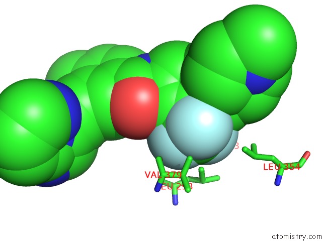
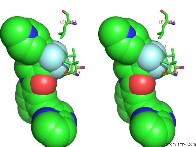
Fluorine binding site 1 out of 3 in 3oxz
Go back to
Fluorine binding site 1 out
of 3 in the Crystal Structure of Abl Kinase Domain Bound with A Dfg-Out Inhibitor AP24534
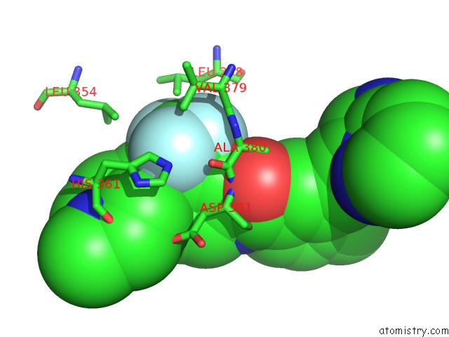
Mono view
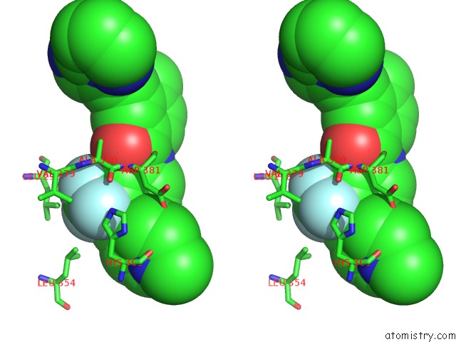
Stereo pair view
Mono view
Stereo pair view
A full contact list of Fluorine with other atoms in the F binding
site number 1 of Crystal Structure of Abl Kinase Domain Bound with A Dfg-Out Inhibitor AP24534 within 5.0Å range:
|
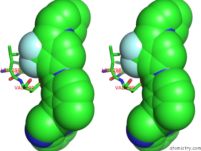
Fluorine binding site 2 out of 3 in 3oxz
Go back to
Fluorine binding site 2 out
of 3 in the Crystal Structure of Abl Kinase Domain Bound with A Dfg-Out Inhibitor AP24534
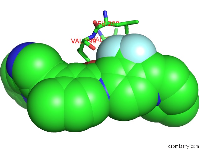
Mono view
Stereo pair view
Mono view
Stereo pair view
A full contact list of Fluorine with other atoms in the F binding
site number 2 of Crystal Structure of Abl Kinase Domain Bound with A Dfg-Out Inhibitor AP24534 within 5.0Å range:
|
Fluorine binding site 3 out of 3 in 3oxz
Go back to
Fluorine binding site 3 out
of 3 in the Crystal Structure of Abl Kinase Domain Bound with A Dfg-Out Inhibitor AP24534
Mono view
Stereo pair view
Mono view
Stereo pair view
A full contact list of Fluorine with other atoms in the F binding
site number 3 of Crystal Structure of Abl Kinase Domain Bound with A Dfg-Out Inhibitor AP24534 within 5.0Å range:
|
Reference:
T.Zhou,
L.Commodore,
W.S.Huang,
Y.Wang,
M.Thomas,
J.Keats,
Q.Xu,
V.M.Rivera,
W.C.Shakespeare,
T.Clackson,
D.C.Dalgarno,
X.Zhu.
Structural Mechanism of the Pan-Bcr-Abl Inhibitor Ponatinib (AP24534): Lessons For Overcoming Kinase Inhibitor Resistance. Chem.Biol.Drug Des. V. 77 1 2011.
ISSN: ISSN 1747-0277
PubMed: 21118377
DOI: 10.1111/J.1747-0285.2010.01054.X
Page generated: Wed Jul 31 21:28:55 2024
ISSN: ISSN 1747-0277
PubMed: 21118377
DOI: 10.1111/J.1747-0285.2010.01054.X
Last articles
Zn in 9J0NZn in 9J0O
Zn in 9J0P
Zn in 9FJX
Zn in 9EKB
Zn in 9C0F
Zn in 9CAH
Zn in 9CH0
Zn in 9CH3
Zn in 9CH1