Fluorine »
PDB 3oyl-3pr2 »
3p5s »
Fluorine in PDB 3p5s: Structural Insights Into the Catalytic Mechanism of CD38: Evidence For A Conformationally Flexible Covalent Enzyme-Substrate Complex
Enzymatic activity of Structural Insights Into the Catalytic Mechanism of CD38: Evidence For A Conformationally Flexible Covalent Enzyme-Substrate Complex
All present enzymatic activity of Structural Insights Into the Catalytic Mechanism of CD38: Evidence For A Conformationally Flexible Covalent Enzyme-Substrate Complex:
3.2.2.5;
3.2.2.5;
Protein crystallography data
The structure of Structural Insights Into the Catalytic Mechanism of CD38: Evidence For A Conformationally Flexible Covalent Enzyme-Substrate Complex, PDB code: 3p5s
was solved by
P.F.Egea,
H.Muller-Stauffler,
I.Kohn,
C.Cakou-Kefir,
R.M.Stroud,
E.Kellenberburger,
F.Schuber,
with X-Ray Crystallography technique. A brief refinement statistics is given in the table below:
| Resolution Low / High (Å) | 40.01 / 1.95 |
| Space group | P 21 21 21 |
| Cell size a, b, c (Å), α, β, γ (°) | 46.205, 80.029, 157.725, 90.00, 90.00, 90.00 |
| R / Rfree (%) | 20.5 / 25.2 |
Fluorine Binding Sites:
The binding sites of Fluorine atom in the Structural Insights Into the Catalytic Mechanism of CD38: Evidence For A Conformationally Flexible Covalent Enzyme-Substrate Complex
(pdb code 3p5s). This binding sites where shown within
5.0 Angstroms radius around Fluorine atom.
In total 2 binding sites of Fluorine where determined in the Structural Insights Into the Catalytic Mechanism of CD38: Evidence For A Conformationally Flexible Covalent Enzyme-Substrate Complex, PDB code: 3p5s:
Jump to Fluorine binding site number: 1; 2;
In total 2 binding sites of Fluorine where determined in the Structural Insights Into the Catalytic Mechanism of CD38: Evidence For A Conformationally Flexible Covalent Enzyme-Substrate Complex, PDB code: 3p5s:
Jump to Fluorine binding site number: 1; 2;
Fluorine binding site 1 out of 2 in 3p5s
Go back to
Fluorine binding site 1 out
of 2 in the Structural Insights Into the Catalytic Mechanism of CD38: Evidence For A Conformationally Flexible Covalent Enzyme-Substrate Complex
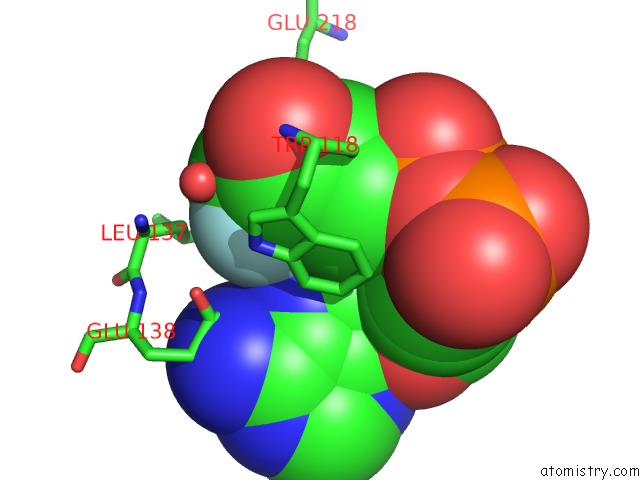
Mono view
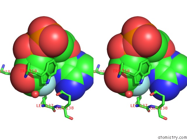
Stereo pair view
Mono view
Stereo pair view
A full contact list of Fluorine with other atoms in the F binding
site number 1 of Structural Insights Into the Catalytic Mechanism of CD38: Evidence For A Conformationally Flexible Covalent Enzyme-Substrate Complex within 5.0Å range:
|
Fluorine binding site 2 out of 2 in 3p5s
Go back to
Fluorine binding site 2 out
of 2 in the Structural Insights Into the Catalytic Mechanism of CD38: Evidence For A Conformationally Flexible Covalent Enzyme-Substrate Complex
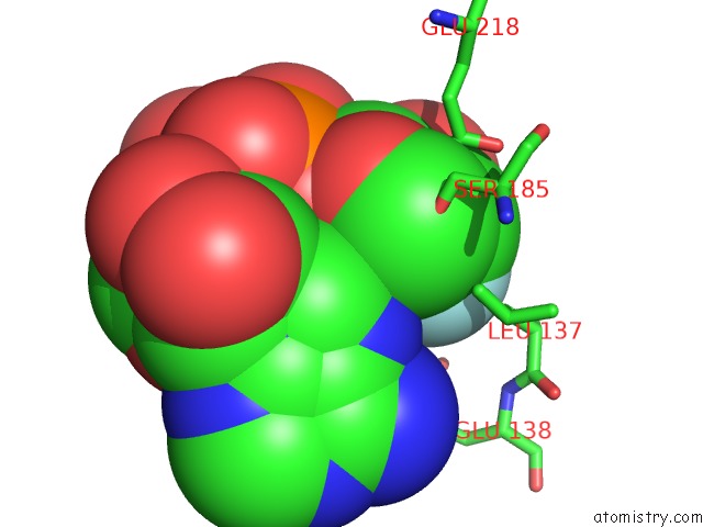
Mono view
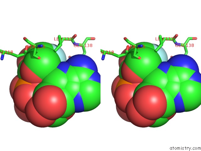
Stereo pair view
Mono view
Stereo pair view
A full contact list of Fluorine with other atoms in the F binding
site number 2 of Structural Insights Into the Catalytic Mechanism of CD38: Evidence For A Conformationally Flexible Covalent Enzyme-Substrate Complex within 5.0Å range:
|
Reference:
P.F.Egea,
H.Muller-Steffner,
I.Kuhn,
C.Cakir-Kiefer,
N.J.Oppenheimer,
R.M.Stroud,
E.Kellenberger,
F.Schuber.
Insights Into the Mechanism of Bovine CD38/Nad+Glycohydrolase From the X-Ray Structures of Its Michaelis Complex and Covalently-Trapped Intermediates. Plos One V. 7 34918 2012.
ISSN: ESSN 1932-6203
PubMed: 22529956
DOI: 10.1371/JOURNAL.PONE.0034918
Page generated: Wed Jul 31 21:38:19 2024
ISSN: ESSN 1932-6203
PubMed: 22529956
DOI: 10.1371/JOURNAL.PONE.0034918
Last articles
Zn in 9J0NZn in 9J0O
Zn in 9J0P
Zn in 9FJX
Zn in 9EKB
Zn in 9C0F
Zn in 9CAH
Zn in 9CH0
Zn in 9CH3
Zn in 9CH1