Fluorine »
PDB 3oyl-3pr2 »
3pp0 »
Fluorine in PDB 3pp0: Crystal Structure of the Kinase Domain of Human HER2 (ERBB2).
Enzymatic activity of Crystal Structure of the Kinase Domain of Human HER2 (ERBB2).
All present enzymatic activity of Crystal Structure of the Kinase Domain of Human HER2 (ERBB2).:
2.7.10.1;
2.7.10.1;
Protein crystallography data
The structure of Crystal Structure of the Kinase Domain of Human HER2 (ERBB2)., PDB code: 3pp0
was solved by
R.J.Skene,
K.Aertgeerts,
S.Sogabe,
with X-Ray Crystallography technique. A brief refinement statistics is given in the table below:
| Resolution Low / High (Å) | 50.00 / 2.25 |
| Space group | P 21 21 21 |
| Cell size a, b, c (Å), α, β, γ (°) | 48.705, 78.951, 152.675, 90.00, 90.00, 90.00 |
| R / Rfree (%) | 18.5 / 26 |
Other elements in 3pp0:
The structure of Crystal Structure of the Kinase Domain of Human HER2 (ERBB2). also contains other interesting chemical elements:
| Chlorine | (Cl) | 2 atoms |
Fluorine Binding Sites:
The binding sites of Fluorine atom in the Crystal Structure of the Kinase Domain of Human HER2 (ERBB2).
(pdb code 3pp0). This binding sites where shown within
5.0 Angstroms radius around Fluorine atom.
In total 6 binding sites of Fluorine where determined in the Crystal Structure of the Kinase Domain of Human HER2 (ERBB2)., PDB code: 3pp0:
Jump to Fluorine binding site number: 1; 2; 3; 4; 5; 6;
In total 6 binding sites of Fluorine where determined in the Crystal Structure of the Kinase Domain of Human HER2 (ERBB2)., PDB code: 3pp0:
Jump to Fluorine binding site number: 1; 2; 3; 4; 5; 6;
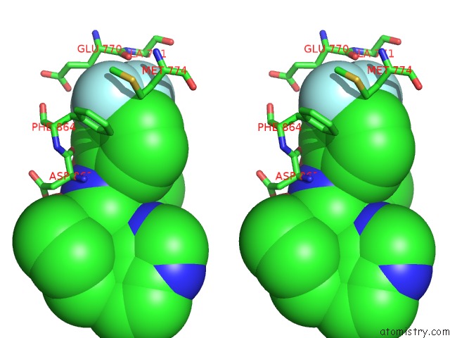
Fluorine binding site 1 out of 6 in 3pp0
Go back to
Fluorine binding site 1 out
of 6 in the Crystal Structure of the Kinase Domain of Human HER2 (ERBB2).
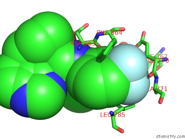
Mono view
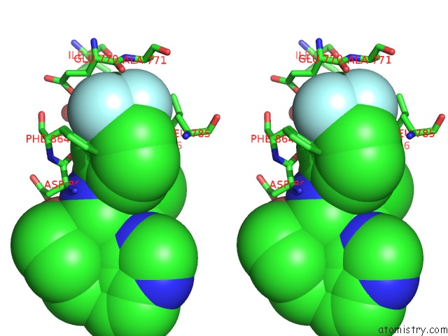
Stereo pair view
Mono view
Stereo pair view
A full contact list of Fluorine with other atoms in the F binding
site number 1 of Crystal Structure of the Kinase Domain of Human HER2 (ERBB2). within 5.0Å range:
|
Fluorine binding site 2 out of 6 in 3pp0
Go back to
Fluorine binding site 2 out
of 6 in the Crystal Structure of the Kinase Domain of Human HER2 (ERBB2).
Mono view

Stereo pair view
Mono view
Stereo pair view
A full contact list of Fluorine with other atoms in the F binding
site number 2 of Crystal Structure of the Kinase Domain of Human HER2 (ERBB2). within 5.0Å range:
|
Fluorine binding site 3 out of 6 in 3pp0
Go back to
Fluorine binding site 3 out
of 6 in the Crystal Structure of the Kinase Domain of Human HER2 (ERBB2).
Mono view
Stereo pair view
Mono view
Stereo pair view
A full contact list of Fluorine with other atoms in the F binding
site number 3 of Crystal Structure of the Kinase Domain of Human HER2 (ERBB2). within 5.0Å range:
|
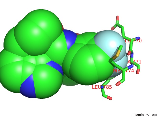
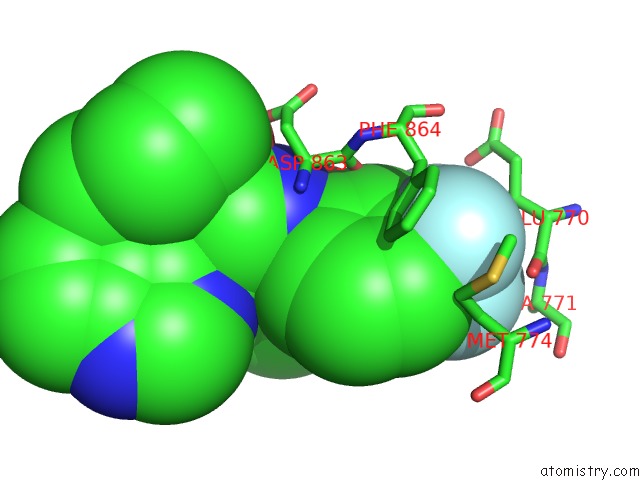
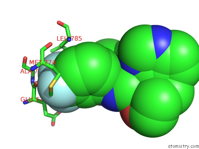
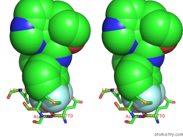
Fluorine binding site 4 out of 6 in 3pp0
Go back to
Fluorine binding site 4 out
of 6 in the Crystal Structure of the Kinase Domain of Human HER2 (ERBB2).

Mono view

Stereo pair view
Mono view
Stereo pair view
A full contact list of Fluorine with other atoms in the F binding
site number 4 of Crystal Structure of the Kinase Domain of Human HER2 (ERBB2). within 5.0Å range:
|
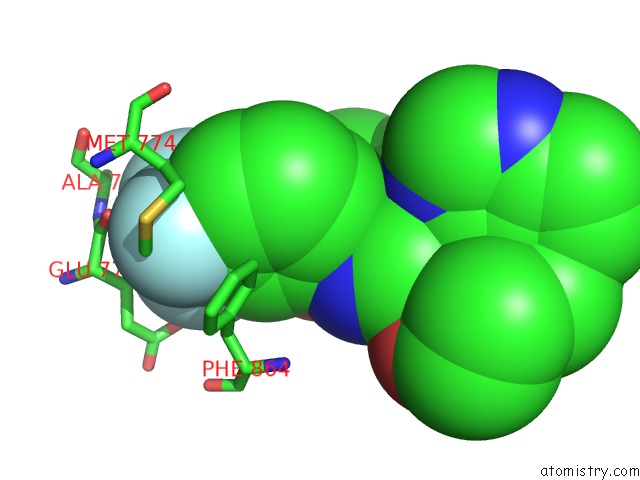
Fluorine binding site 5 out of 6 in 3pp0
Go back to
Fluorine binding site 5 out
of 6 in the Crystal Structure of the Kinase Domain of Human HER2 (ERBB2).
Mono view
Stereo pair view
Mono view
Stereo pair view
A full contact list of Fluorine with other atoms in the F binding
site number 5 of Crystal Structure of the Kinase Domain of Human HER2 (ERBB2). within 5.0Å range:
|
Fluorine binding site 6 out of 6 in 3pp0
Go back to
Fluorine binding site 6 out
of 6 in the Crystal Structure of the Kinase Domain of Human HER2 (ERBB2).
Mono view

Stereo pair view
Mono view
Stereo pair view
A full contact list of Fluorine with other atoms in the F binding
site number 6 of Crystal Structure of the Kinase Domain of Human HER2 (ERBB2). within 5.0Å range:
|
Reference:
K.Aertgeerts,
R.Skene,
J.Yano,
B.C.Sang,
H.Zou,
G.Snell,
A.Jennings,
K.Iwamoto,
N.Habuka,
A.Hirokawa,
T.Ishikawa,
T.Tanaka,
H.Miki,
Y.Ohta,
S.Sogabe.
Structural Analysis of the Mechanism of Inhibition and Allosteric Activation of the Kinase Domain of HER2 Protein. J.Biol.Chem. V. 286 18756 2011.
ISSN: ISSN 0021-9258
PubMed: 21454582
DOI: 10.1074/JBC.M110.206193
Page generated: Wed Jul 31 21:46:23 2024
ISSN: ISSN 0021-9258
PubMed: 21454582
DOI: 10.1074/JBC.M110.206193
Last articles
Zn in 9J0NZn in 9J0O
Zn in 9J0P
Zn in 9FJX
Zn in 9EKB
Zn in 9C0F
Zn in 9CAH
Zn in 9CH0
Zn in 9CH3
Zn in 9CH1