Fluorine »
PDB 3qrj-3rgf »
3rgf »
Fluorine in PDB 3rgf: Crystal Structure of Human CDK8/Cycc
Enzymatic activity of Crystal Structure of Human CDK8/Cycc
Protein crystallography data
The structure of Crystal Structure of Human CDK8/Cycc, PDB code: 3rgf
was solved by
E.V.Schneider,
J.Boettcher,
M.Blaesse,
R.Huber,
K.Maskos,
with X-Ray Crystallography technique. A brief refinement statistics is given in the table below:
| Resolution Low / High (Å) | 37.57 / 2.20 |
| Space group | P 1 21 1 |
| Cell size a, b, c (Å), α, β, γ (°) | 70.690, 70.610, 79.140, 90.00, 108.31, 90.00 |
| R / Rfree (%) | 18 / 22.1 |
Other elements in 3rgf:
The structure of Crystal Structure of Human CDK8/Cycc also contains other interesting chemical elements:
| Chlorine | (Cl) | 1 atom |
Fluorine Binding Sites:
The binding sites of Fluorine atom in the Crystal Structure of Human CDK8/Cycc
(pdb code 3rgf). This binding sites where shown within
5.0 Angstroms radius around Fluorine atom.
In total 3 binding sites of Fluorine where determined in the Crystal Structure of Human CDK8/Cycc, PDB code: 3rgf:
Jump to Fluorine binding site number: 1; 2; 3;
In total 3 binding sites of Fluorine where determined in the Crystal Structure of Human CDK8/Cycc, PDB code: 3rgf:
Jump to Fluorine binding site number: 1; 2; 3;
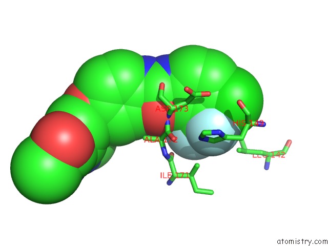
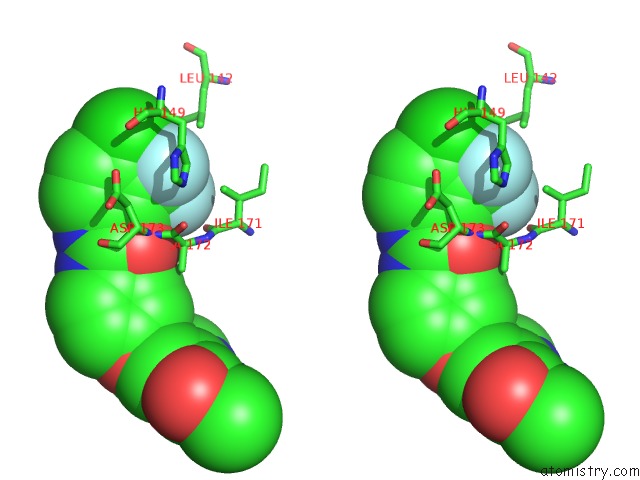
Fluorine binding site 1 out of 3 in 3rgf
Go back to
Fluorine binding site 1 out
of 3 in the Crystal Structure of Human CDK8/Cycc
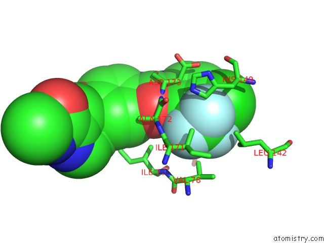
Mono view
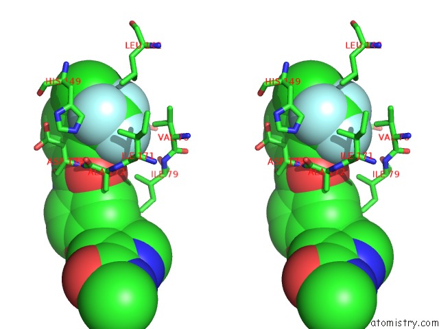
Stereo pair view
Mono view
Stereo pair view
A full contact list of Fluorine with other atoms in the F binding
site number 1 of Crystal Structure of Human CDK8/Cycc within 5.0Å range:
|
Fluorine binding site 2 out of 3 in 3rgf
Go back to
Fluorine binding site 2 out
of 3 in the Crystal Structure of Human CDK8/Cycc
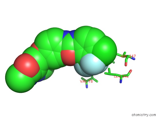
Mono view
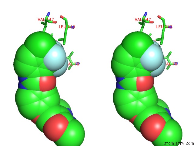
Stereo pair view
Mono view
Stereo pair view
A full contact list of Fluorine with other atoms in the F binding
site number 2 of Crystal Structure of Human CDK8/Cycc within 5.0Å range:
|
Fluorine binding site 3 out of 3 in 3rgf
Go back to
Fluorine binding site 3 out
of 3 in the Crystal Structure of Human CDK8/Cycc
Mono view
Stereo pair view
Mono view
Stereo pair view
A full contact list of Fluorine with other atoms in the F binding
site number 3 of Crystal Structure of Human CDK8/Cycc within 5.0Å range:
|
Reference:
E.V.Schneider,
J.Bottcher,
M.Blaesse,
L.Neumann,
R.Huber,
K.Maskos.
The Structure of CDK8/Cycc Implicates Specificity in the Cdk/Cyclin Family and Reveals Interaction with A Deep Pocket Binder. J.Mol.Biol. V. 412 251 2011.
ISSN: ISSN 0022-2836
PubMed: 21806996
DOI: 10.1016/J.JMB.2011.07.020
Page generated: Wed Jul 31 22:16:17 2024
ISSN: ISSN 0022-2836
PubMed: 21806996
DOI: 10.1016/J.JMB.2011.07.020
Last articles
Zn in 9J0NZn in 9J0O
Zn in 9J0P
Zn in 9FJX
Zn in 9EKB
Zn in 9C0F
Zn in 9CAH
Zn in 9CH0
Zn in 9CH3
Zn in 9CH1