Fluorine »
PDB 3rjc-3s9y »
3rlj »
Fluorine in PDB 3rlj: Crystal Structure of the Androgen Receptor Ligand Binding Domain in Complex with Sarm S-22
Protein crystallography data
The structure of Crystal Structure of the Androgen Receptor Ligand Binding Domain in Complex with Sarm S-22, PDB code: 3rlj
was solved by
C.E.Bohl,
C.B.Duke,
A.Jones,
J.T.Dalton,
D.D.Miller,
with X-Ray Crystallography technique. A brief refinement statistics is given in the table below:
| Resolution Low / High (Å) | 23.79 / 1.90 |
| Space group | P 21 21 21 |
| Cell size a, b, c (Å), α, β, γ (°) | 54.742, 66.795, 69.374, 90.00, 90.00, 90.00 |
| R / Rfree (%) | 22.1 / 26.5 |
Fluorine Binding Sites:
The binding sites of Fluorine atom in the Crystal Structure of the Androgen Receptor Ligand Binding Domain in Complex with Sarm S-22
(pdb code 3rlj). This binding sites where shown within
5.0 Angstroms radius around Fluorine atom.
In total 3 binding sites of Fluorine where determined in the Crystal Structure of the Androgen Receptor Ligand Binding Domain in Complex with Sarm S-22, PDB code: 3rlj:
Jump to Fluorine binding site number: 1; 2; 3;
In total 3 binding sites of Fluorine where determined in the Crystal Structure of the Androgen Receptor Ligand Binding Domain in Complex with Sarm S-22, PDB code: 3rlj:
Jump to Fluorine binding site number: 1; 2; 3;
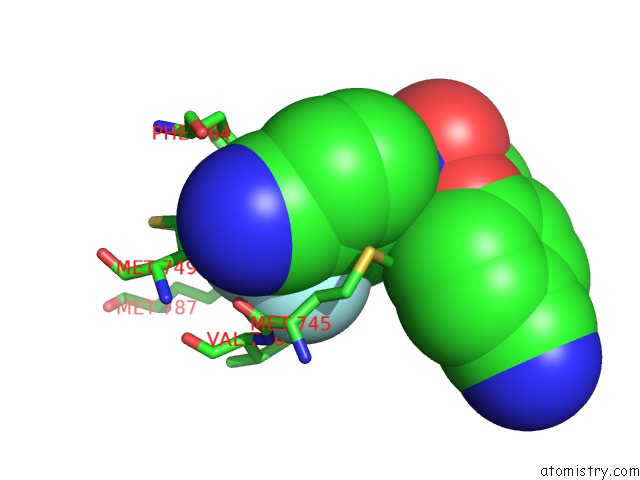
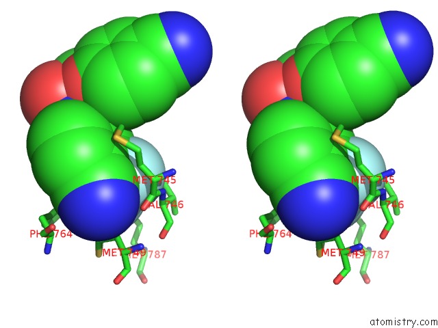
Fluorine binding site 1 out of 3 in 3rlj
Go back to
Fluorine binding site 1 out
of 3 in the Crystal Structure of the Androgen Receptor Ligand Binding Domain in Complex with Sarm S-22
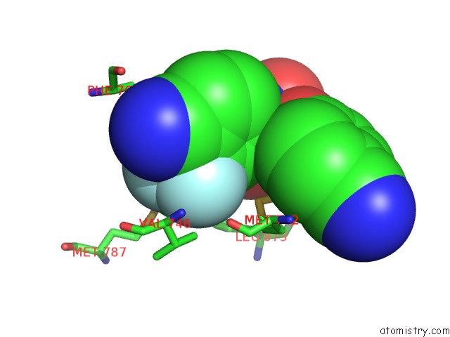
Mono view
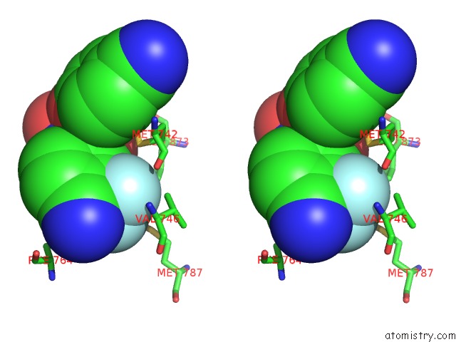
Stereo pair view
Mono view
Stereo pair view
A full contact list of Fluorine with other atoms in the F binding
site number 1 of Crystal Structure of the Androgen Receptor Ligand Binding Domain in Complex with Sarm S-22 within 5.0Å range:
|
Fluorine binding site 2 out of 3 in 3rlj
Go back to
Fluorine binding site 2 out
of 3 in the Crystal Structure of the Androgen Receptor Ligand Binding Domain in Complex with Sarm S-22
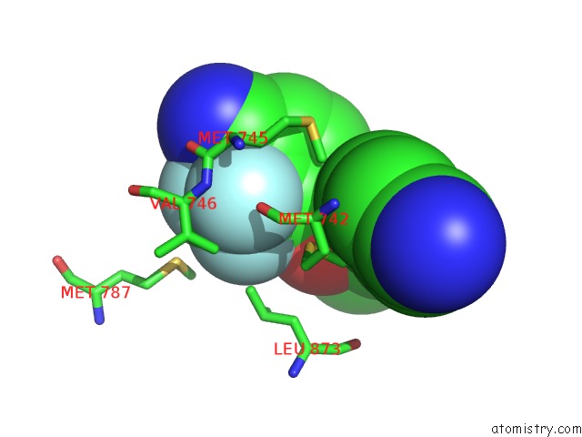
Mono view
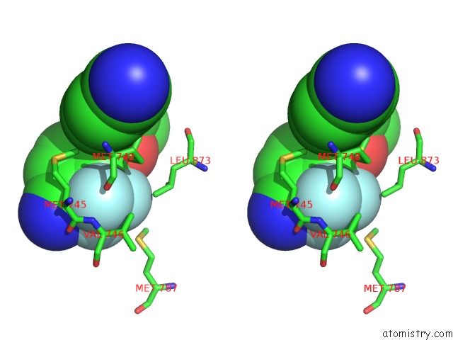
Stereo pair view
Mono view
Stereo pair view
A full contact list of Fluorine with other atoms in the F binding
site number 2 of Crystal Structure of the Androgen Receptor Ligand Binding Domain in Complex with Sarm S-22 within 5.0Å range:
|
Fluorine binding site 3 out of 3 in 3rlj
Go back to
Fluorine binding site 3 out
of 3 in the Crystal Structure of the Androgen Receptor Ligand Binding Domain in Complex with Sarm S-22
Mono view
Stereo pair view
Mono view
Stereo pair view
A full contact list of Fluorine with other atoms in the F binding
site number 3 of Crystal Structure of the Androgen Receptor Ligand Binding Domain in Complex with Sarm S-22 within 5.0Å range:
|
Reference:
C.B.Duke,
A.Jones,
C.E.Bohl,
J.T.Dalton,
D.D.Miller.
Unexpected Binding Orientation of Bulky-B-Ring Anti-Androgens and Implications For Future Drug Targets. J.Med.Chem. V. 54 3973 2011.
ISSN: ISSN 0022-2623
PubMed: 21506597
DOI: 10.1021/JM2000097
Page generated: Mon Jul 14 19:08:51 2025
ISSN: ISSN 0022-2623
PubMed: 21506597
DOI: 10.1021/JM2000097
Last articles
Fe in 2YXOFe in 2YRS
Fe in 2YXC
Fe in 2YNM
Fe in 2YVJ
Fe in 2YP1
Fe in 2YU2
Fe in 2YU1
Fe in 2YQB
Fe in 2YOO