Fluorine »
PDB 3sb0-3sym »
3scq »
Fluorine in PDB 3scq: Crystal Structure of Rice BGLU1 E386A Mutant Complexed with Alpha- Glucosyl Fluoride
Enzymatic activity of Crystal Structure of Rice BGLU1 E386A Mutant Complexed with Alpha- Glucosyl Fluoride
All present enzymatic activity of Crystal Structure of Rice BGLU1 E386A Mutant Complexed with Alpha- Glucosyl Fluoride:
3.2.1.21;
3.2.1.21;
Protein crystallography data
The structure of Crystal Structure of Rice BGLU1 E386A Mutant Complexed with Alpha- Glucosyl Fluoride, PDB code: 3scq
was solved by
S.Pengthaisong,
S.G.Withers,
B.Kuaprasert,
J.R.Ketudat Cairns,
with X-Ray Crystallography technique. A brief refinement statistics is given in the table below:
| Resolution Low / High (Å) | 25.01 / 2.10 |
| Space group | P 21 21 21 |
| Cell size a, b, c (Å), α, β, γ (°) | 80.117, 100.905, 126.828, 90.00, 90.00, 90.00 |
| R / Rfree (%) | 17.5 / 20.6 |
Other elements in 3scq:
The structure of Crystal Structure of Rice BGLU1 E386A Mutant Complexed with Alpha- Glucosyl Fluoride also contains other interesting chemical elements:
| Zinc | (Zn) | 1 atom |
Fluorine Binding Sites:
The binding sites of Fluorine atom in the Crystal Structure of Rice BGLU1 E386A Mutant Complexed with Alpha- Glucosyl Fluoride
(pdb code 3scq). This binding sites where shown within
5.0 Angstroms radius around Fluorine atom.
In total 2 binding sites of Fluorine where determined in the Crystal Structure of Rice BGLU1 E386A Mutant Complexed with Alpha- Glucosyl Fluoride, PDB code: 3scq:
Jump to Fluorine binding site number: 1; 2;
In total 2 binding sites of Fluorine where determined in the Crystal Structure of Rice BGLU1 E386A Mutant Complexed with Alpha- Glucosyl Fluoride, PDB code: 3scq:
Jump to Fluorine binding site number: 1; 2;
Fluorine binding site 1 out of 2 in 3scq
Go back to
Fluorine binding site 1 out
of 2 in the Crystal Structure of Rice BGLU1 E386A Mutant Complexed with Alpha- Glucosyl Fluoride
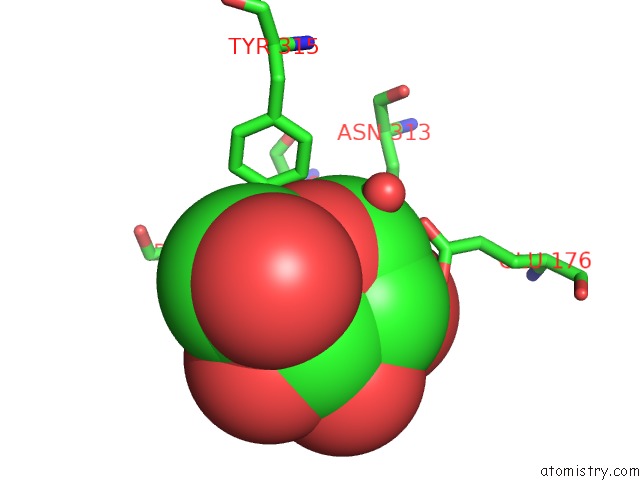
Mono view
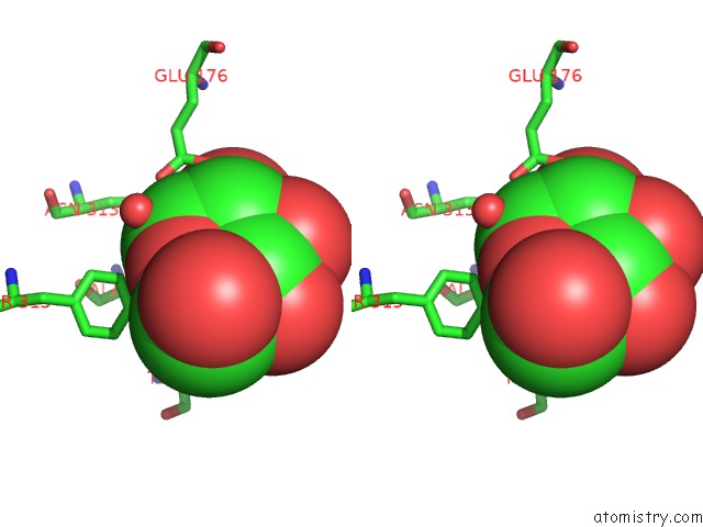
Stereo pair view
Mono view
Stereo pair view
A full contact list of Fluorine with other atoms in the F binding
site number 1 of Crystal Structure of Rice BGLU1 E386A Mutant Complexed with Alpha- Glucosyl Fluoride within 5.0Å range:
|
Fluorine binding site 2 out of 2 in 3scq
Go back to
Fluorine binding site 2 out
of 2 in the Crystal Structure of Rice BGLU1 E386A Mutant Complexed with Alpha- Glucosyl Fluoride
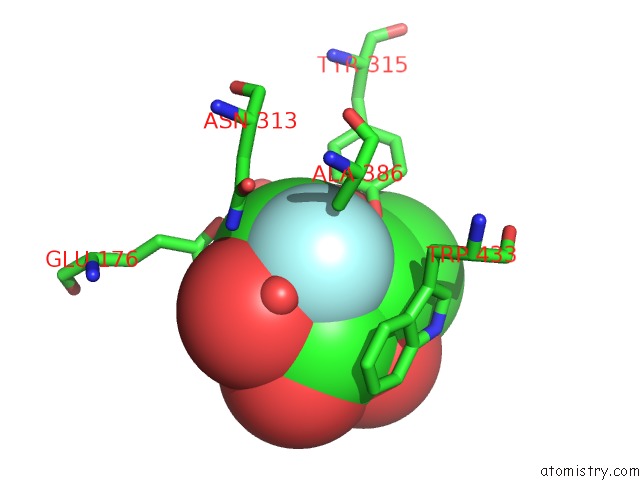
Mono view
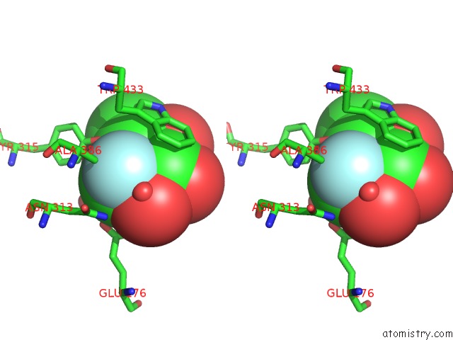
Stereo pair view
Mono view
Stereo pair view
A full contact list of Fluorine with other atoms in the F binding
site number 2 of Crystal Structure of Rice BGLU1 E386A Mutant Complexed with Alpha- Glucosyl Fluoride within 5.0Å range:
|
Reference:
S.Pengthaisong,
S.G.Withers,
B.Kuaprasert,
J.R.Ketudat Cairns.
Structural Investigation of the Basis For Cellooligosaccharide Synthesis By Rice BGLU1 Glycosynthases To Be Published.
Page generated: Mon Jul 14 19:16:51 2025
Last articles
Fe in 2YXOFe in 2YRS
Fe in 2YXC
Fe in 2YNM
Fe in 2YVJ
Fe in 2YP1
Fe in 2YU2
Fe in 2YU1
Fe in 2YQB
Fe in 2YOO