Fluorine »
PDB 3sb0-3sym »
3sji »
Fluorine in PDB 3sji: Crystal Structure of CVA16 3C in Complex with Rupintrivir (AG7088)
Enzymatic activity of Crystal Structure of CVA16 3C in Complex with Rupintrivir (AG7088)
All present enzymatic activity of Crystal Structure of CVA16 3C in Complex with Rupintrivir (AG7088):
3.4.22.28;
3.4.22.28;
Protein crystallography data
The structure of Crystal Structure of CVA16 3C in Complex with Rupintrivir (AG7088), PDB code: 3sji
was solved by
G.Lu,
J.Qi,
Z.Chen,
X.Xu,
F.Gao,
D.Lin,
W.Qian,
H.Liu,
H.Jiang,
J.Yan,
G.F.Gao,
with X-Ray Crystallography technique. A brief refinement statistics is given in the table below:
| Resolution Low / High (Å) | 28.57 / 1.80 |
| Space group | P 43 |
| Cell size a, b, c (Å), α, β, γ (°) | 40.406, 40.406, 98.740, 90.00, 90.00, 90.00 |
| R / Rfree (%) | 17.9 / 21.8 |
Other elements in 3sji:
The structure of Crystal Structure of CVA16 3C in Complex with Rupintrivir (AG7088) also contains other interesting chemical elements:
| Sodium | (Na) | 1 atom |
Fluorine Binding Sites:
The binding sites of Fluorine atom in the Crystal Structure of CVA16 3C in Complex with Rupintrivir (AG7088)
(pdb code 3sji). This binding sites where shown within
5.0 Angstroms radius around Fluorine atom.
In total only one binding site of Fluorine was determined in the Crystal Structure of CVA16 3C in Complex with Rupintrivir (AG7088), PDB code: 3sji:
In total only one binding site of Fluorine was determined in the Crystal Structure of CVA16 3C in Complex with Rupintrivir (AG7088), PDB code: 3sji:
Fluorine binding site 1 out of 1 in 3sji
Go back to
Fluorine binding site 1 out
of 1 in the Crystal Structure of CVA16 3C in Complex with Rupintrivir (AG7088)
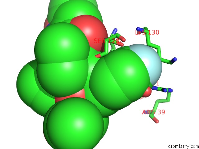
Mono view
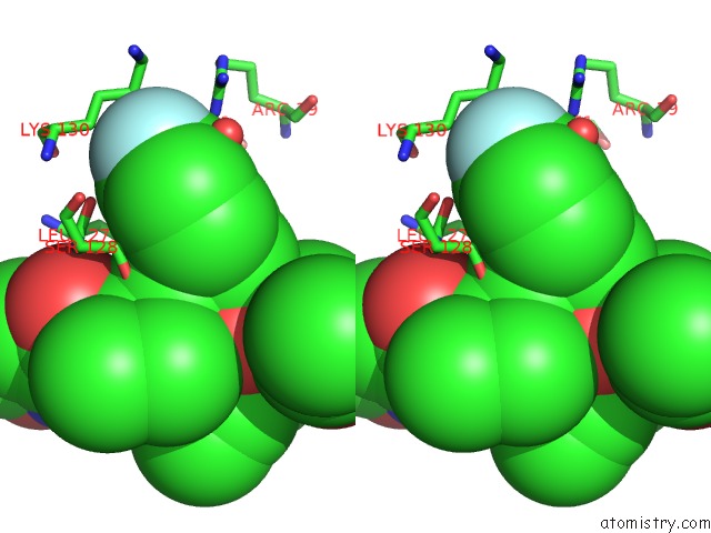
Stereo pair view
Mono view
Stereo pair view
A full contact list of Fluorine with other atoms in the F binding
site number 1 of Crystal Structure of CVA16 3C in Complex with Rupintrivir (AG7088) within 5.0Å range:
|
Reference:
G.Lu,
J.Qi,
Z.Chen,
X.Xu,
F.Gao,
D.Lin,
W.Qian,
H.Liu,
H.Jiang,
J.Yan,
G.F.Gao.
Enterovirus 71 and Coxsackievirus A16 3C Proteases: Binding to Rupintrivir and Their Substrates and Anti-Hand, Foot, and Mouth Disease Virus Drug Design. J.Virol. V. 85 10319 2011.
ISSN: ISSN 0022-538X
PubMed: 21795339
DOI: 10.1128/JVI.00787-11
Page generated: Wed Jul 31 22:32:31 2024
ISSN: ISSN 0022-538X
PubMed: 21795339
DOI: 10.1128/JVI.00787-11
Last articles
Zn in 9J0NZn in 9J0O
Zn in 9J0P
Zn in 9FJX
Zn in 9EKB
Zn in 9C0F
Zn in 9CAH
Zn in 9CH0
Zn in 9CH3
Zn in 9CH1