Fluorine »
PDB 3sb0-3sym »
3spk »
Fluorine in PDB 3spk: Tipranavir in Complex with A Human Immunodeficiency Virus Type 1 Protease Variant
Enzymatic activity of Tipranavir in Complex with A Human Immunodeficiency Virus Type 1 Protease Variant
All present enzymatic activity of Tipranavir in Complex with A Human Immunodeficiency Virus Type 1 Protease Variant:
3.4.23.16;
3.4.23.16;
Protein crystallography data
The structure of Tipranavir in Complex with A Human Immunodeficiency Virus Type 1 Protease Variant, PDB code: 3spk
was solved by
Y.Wang,
Z.Liu,
J.S.Brunzelle,
I.A.Kovari,
L.C.Kovari,
with X-Ray Crystallography technique. A brief refinement statistics is given in the table below:
| Resolution Low / High (Å) | 29.52 / 1.24 |
| Space group | P 61 |
| Cell size a, b, c (Å), α, β, γ (°) | 63.119, 63.119, 83.540, 90.00, 90.00, 120.00 |
| R / Rfree (%) | 17.4 / 22.7 |
Fluorine Binding Sites:
The binding sites of Fluorine atom in the Tipranavir in Complex with A Human Immunodeficiency Virus Type 1 Protease Variant
(pdb code 3spk). This binding sites where shown within
5.0 Angstroms radius around Fluorine atom.
In total 6 binding sites of Fluorine where determined in the Tipranavir in Complex with A Human Immunodeficiency Virus Type 1 Protease Variant, PDB code: 3spk:
Jump to Fluorine binding site number: 1; 2; 3; 4; 5; 6;
In total 6 binding sites of Fluorine where determined in the Tipranavir in Complex with A Human Immunodeficiency Virus Type 1 Protease Variant, PDB code: 3spk:
Jump to Fluorine binding site number: 1; 2; 3; 4; 5; 6;
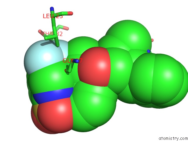
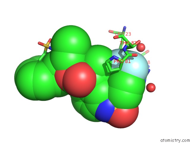
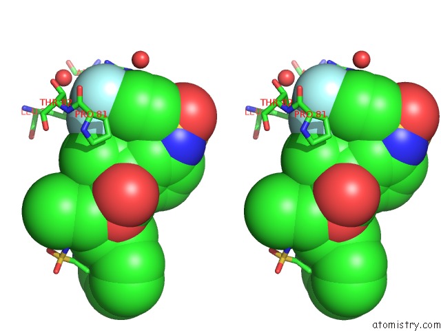
Fluorine binding site 1 out of 6 in 3spk
Go back to
Fluorine binding site 1 out
of 6 in the Tipranavir in Complex with A Human Immunodeficiency Virus Type 1 Protease Variant
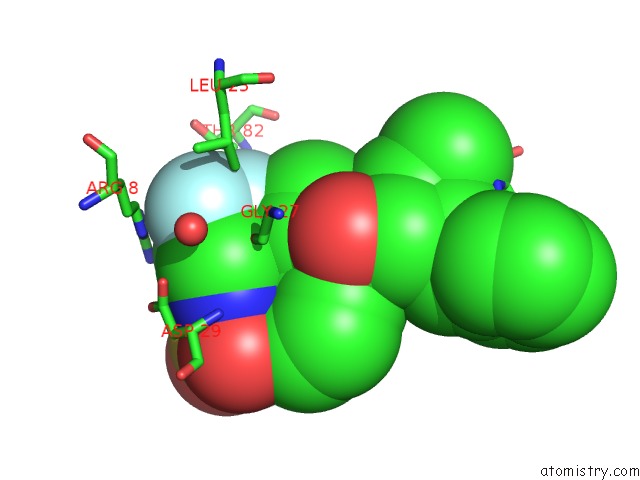
Mono view
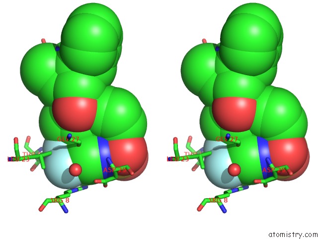
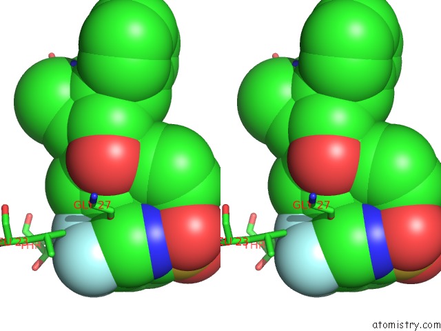
Stereo pair view
Mono view
Stereo pair view
A full contact list of Fluorine with other atoms in the F binding
site number 1 of Tipranavir in Complex with A Human Immunodeficiency Virus Type 1 Protease Variant within 5.0Å range:
|
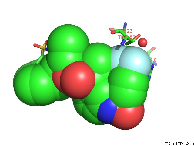
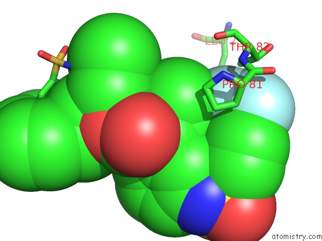
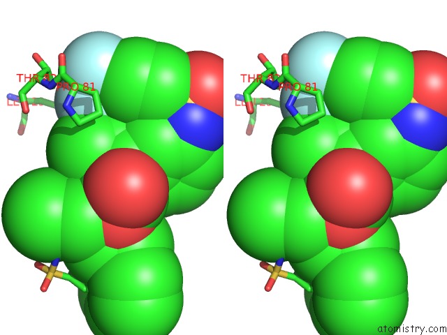
Fluorine binding site 2 out of 6 in 3spk
Go back to
Fluorine binding site 2 out
of 6 in the Tipranavir in Complex with A Human Immunodeficiency Virus Type 1 Protease Variant
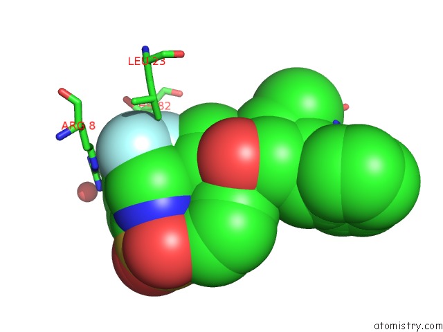
Mono view
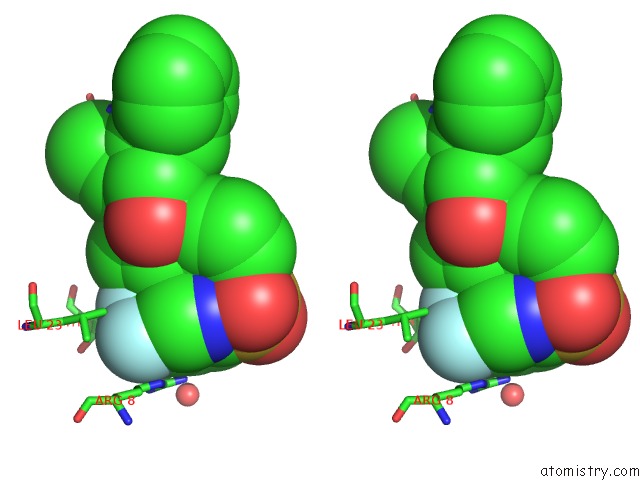
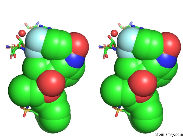
Stereo pair view
Mono view
Stereo pair view
A full contact list of Fluorine with other atoms in the F binding
site number 2 of Tipranavir in Complex with A Human Immunodeficiency Virus Type 1 Protease Variant within 5.0Å range:
|
Fluorine binding site 3 out of 6 in 3spk
Go back to
Fluorine binding site 3 out
of 6 in the Tipranavir in Complex with A Human Immunodeficiency Virus Type 1 Protease Variant
Mono view
Stereo pair view
Mono view
Stereo pair view
A full contact list of Fluorine with other atoms in the F binding
site number 3 of Tipranavir in Complex with A Human Immunodeficiency Virus Type 1 Protease Variant within 5.0Å range:
|
Fluorine binding site 4 out of 6 in 3spk
Go back to
Fluorine binding site 4 out
of 6 in the Tipranavir in Complex with A Human Immunodeficiency Virus Type 1 Protease Variant
Mono view
Stereo pair view
Mono view
Stereo pair view
A full contact list of Fluorine with other atoms in the F binding
site number 4 of Tipranavir in Complex with A Human Immunodeficiency Virus Type 1 Protease Variant within 5.0Å range:
|
Fluorine binding site 5 out of 6 in 3spk
Go back to
Fluorine binding site 5 out
of 6 in the Tipranavir in Complex with A Human Immunodeficiency Virus Type 1 Protease Variant
Mono view
Stereo pair view
Mono view
Stereo pair view
A full contact list of Fluorine with other atoms in the F binding
site number 5 of Tipranavir in Complex with A Human Immunodeficiency Virus Type 1 Protease Variant within 5.0Å range:
|
Fluorine binding site 6 out of 6 in 3spk
Go back to
Fluorine binding site 6 out
of 6 in the Tipranavir in Complex with A Human Immunodeficiency Virus Type 1 Protease Variant
Mono view
Stereo pair view
Mono view
Stereo pair view
A full contact list of Fluorine with other atoms in the F binding
site number 6 of Tipranavir in Complex with A Human Immunodeficiency Virus Type 1 Protease Variant within 5.0Å range:
|
Reference:
Y.Wang,
Z.Liu,
J.S.Brunzelle,
I.A.Kovari,
T.G.Dewdney,
S.J.Reiter,
L.C.Kovari.
The Higher Barrier of Darunavir and Tipranavir Resistance For Hiv-1 Protease. Biochem.Biophys.Res.Commun. V. 412 737 2011.
ISSN: ISSN 0006-291X
PubMed: 21871444
DOI: 10.1016/J.BBRC.2011.08.045
Page generated: Wed Jul 31 22:35:59 2024
ISSN: ISSN 0006-291X
PubMed: 21871444
DOI: 10.1016/J.BBRC.2011.08.045
Last articles
Zn in 9JYWZn in 9IR4
Zn in 9IR3
Zn in 9GMX
Zn in 9GMW
Zn in 9JEJ
Zn in 9ERF
Zn in 9ERE
Zn in 9EGV
Zn in 9EGW