Fluorine »
PDB 3syn-3u2o »
3tu1 »
Fluorine in PDB 3tu1: Exhaustive Fluorine Scanning Towards Potent P53-MDM2 Antagonist
Protein crystallography data
The structure of Exhaustive Fluorine Scanning Towards Potent P53-MDM2 Antagonist, PDB code: 3tu1
was solved by
S.Wolf,
Y.Huang,
D.Koes,
G.M.Popowicz,
C.J.Camacho,
T.A.Holak,
A.Doemling,
with X-Ray Crystallography technique. A brief refinement statistics is given in the table below:
| Resolution Low / High (Å) | 20.00 / 1.60 |
| Space group | I 2 2 2 |
| Cell size a, b, c (Å), α, β, γ (°) | 49.950, 59.250, 82.950, 90.00, 90.00, 90.00 |
| R / Rfree (%) | 20.5 / 24.5 |
Other elements in 3tu1:
The structure of Exhaustive Fluorine Scanning Towards Potent P53-MDM2 Antagonist also contains other interesting chemical elements:
| Chlorine | (Cl) | 1 atom |
Fluorine Binding Sites:
The binding sites of Fluorine atom in the Exhaustive Fluorine Scanning Towards Potent P53-MDM2 Antagonist
(pdb code 3tu1). This binding sites where shown within
5.0 Angstroms radius around Fluorine atom.
In total 2 binding sites of Fluorine where determined in the Exhaustive Fluorine Scanning Towards Potent P53-MDM2 Antagonist, PDB code: 3tu1:
Jump to Fluorine binding site number: 1; 2;
In total 2 binding sites of Fluorine where determined in the Exhaustive Fluorine Scanning Towards Potent P53-MDM2 Antagonist, PDB code: 3tu1:
Jump to Fluorine binding site number: 1; 2;
Fluorine binding site 1 out of 2 in 3tu1
Go back to
Fluorine binding site 1 out
of 2 in the Exhaustive Fluorine Scanning Towards Potent P53-MDM2 Antagonist
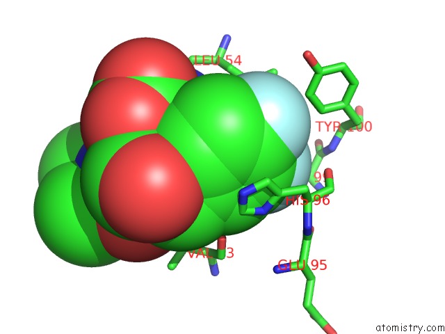
Mono view
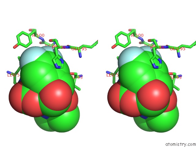
Stereo pair view
Mono view
Stereo pair view
A full contact list of Fluorine with other atoms in the F binding
site number 1 of Exhaustive Fluorine Scanning Towards Potent P53-MDM2 Antagonist within 5.0Å range:
|
Fluorine binding site 2 out of 2 in 3tu1
Go back to
Fluorine binding site 2 out
of 2 in the Exhaustive Fluorine Scanning Towards Potent P53-MDM2 Antagonist
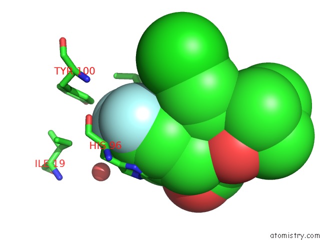
Mono view
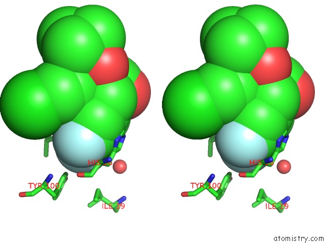
Stereo pair view
Mono view
Stereo pair view
A full contact list of Fluorine with other atoms in the F binding
site number 2 of Exhaustive Fluorine Scanning Towards Potent P53-MDM2 Antagonist within 5.0Å range:
|
Reference:
Y.Huang,
S.Wolf,
D.Koes,
G.M.Popowicz,
C.J.Camacho,
T.A.Holak,
A.Domling.
Exhaustive Fluorine Scanning Toward Potent P53-MDM2 Antagonists. Chemmedchem V. 7 49 2012.
ISSN: ISSN 1860-7179
PubMed: 21954050
DOI: 10.1002/CMDC.201100428
Page generated: Mon Jul 14 19:30:31 2025
ISSN: ISSN 1860-7179
PubMed: 21954050
DOI: 10.1002/CMDC.201100428
Last articles
F in 4EWQF in 4EQU
F in 4EST
F in 4ENH
F in 4EPX
F in 4ENC
F in 4ENB
F in 4EMV
F in 4ENA
F in 4EN5