Fluorine »
PDB 3u5k-3uxm »
3uqw »
Fluorine in PDB 3uqw: Crystal Structure of BACE1 with Its Inhibitor
Enzymatic activity of Crystal Structure of BACE1 with Its Inhibitor
All present enzymatic activity of Crystal Structure of BACE1 with Its Inhibitor:
3.4.23.46;
3.4.23.46;
Protein crystallography data
The structure of Crystal Structure of BACE1 with Its Inhibitor, PDB code: 3uqw
was solved by
T.T.Chen,
W.Y.Chen,
Y.C.Xu,
with X-Ray Crystallography technique. A brief refinement statistics is given in the table below:
| Resolution Low / High (Å) | 36.75 / 2.20 |
| Space group | C 2 2 21 |
| Cell size a, b, c (Å), α, β, γ (°) | 107.315, 131.544, 78.541, 90.00, 90.00, 90.00 |
| R / Rfree (%) | 16.9 / 21.5 |
Fluorine Binding Sites:
The binding sites of Fluorine atom in the Crystal Structure of BACE1 with Its Inhibitor
(pdb code 3uqw). This binding sites where shown within
5.0 Angstroms radius around Fluorine atom.
In total only one binding site of Fluorine was determined in the Crystal Structure of BACE1 with Its Inhibitor, PDB code: 3uqw:
In total only one binding site of Fluorine was determined in the Crystal Structure of BACE1 with Its Inhibitor, PDB code: 3uqw:
Fluorine binding site 1 out of 1 in 3uqw
Go back to
Fluorine binding site 1 out
of 1 in the Crystal Structure of BACE1 with Its Inhibitor
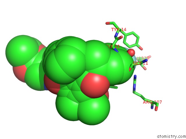
Mono view
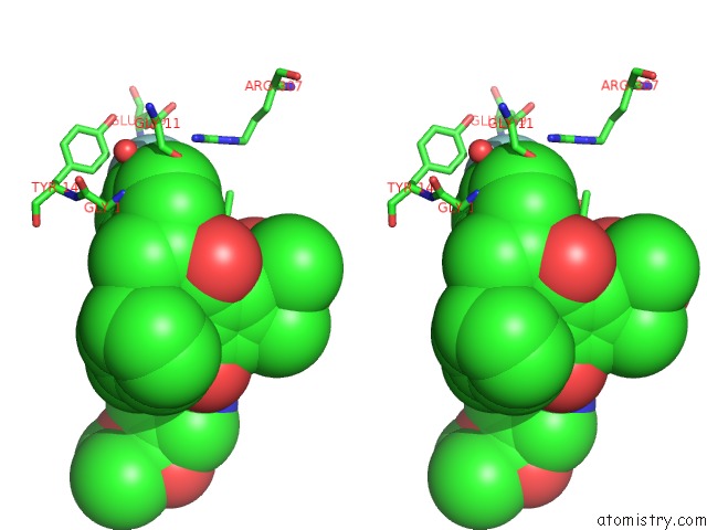
Stereo pair view
Mono view
Stereo pair view
A full contact list of Fluorine with other atoms in the F binding
site number 1 of Crystal Structure of BACE1 with Its Inhibitor within 5.0Å range:
|
Reference:
Y.C.Xu,
W.Y.Chen,
T.T.Chen.
Flexibility of the Flap in the Active Site of BACE1 As Revealed By Crystal Structures and Md Simulations To Be Published.
Page generated: Mon Jul 14 19:46:34 2025
Last articles
F in 4OJRF in 4OI1
F in 4OJ9
F in 4OIU
F in 4OIL
F in 4OHM
F in 4OHK
F in 4OHO
F in 4OI0
F in 4OHA