Fluorine »
PDB 3uyt-3vrb »
3vf6 »
Fluorine in PDB 3vf6: Glucokinase in Complex with Glucose and Activator
Enzymatic activity of Glucokinase in Complex with Glucose and Activator
All present enzymatic activity of Glucokinase in Complex with Glucose and Activator:
2.7.1.2;
2.7.1.2;
Protein crystallography data
The structure of Glucokinase in Complex with Glucose and Activator, PDB code: 3vf6
was solved by
S.Liu,
with X-Ray Crystallography technique. A brief refinement statistics is given in the table below:
| Resolution Low / High (Å) | 52.81 / 1.86 |
| Space group | P 21 21 21 |
| Cell size a, b, c (Å), α, β, γ (°) | 66.850, 82.230, 86.140, 90.00, 90.00, 90.00 |
| R / Rfree (%) | 17.6 / 20.5 |
Other elements in 3vf6:
The structure of Glucokinase in Complex with Glucose and Activator also contains other interesting chemical elements:
| Sodium | (Na) | 1 atom |
Fluorine Binding Sites:
The binding sites of Fluorine atom in the Glucokinase in Complex with Glucose and Activator
(pdb code 3vf6). This binding sites where shown within
5.0 Angstroms radius around Fluorine atom.
In total 3 binding sites of Fluorine where determined in the Glucokinase in Complex with Glucose and Activator, PDB code: 3vf6:
Jump to Fluorine binding site number: 1; 2; 3;
In total 3 binding sites of Fluorine where determined in the Glucokinase in Complex with Glucose and Activator, PDB code: 3vf6:
Jump to Fluorine binding site number: 1; 2; 3;
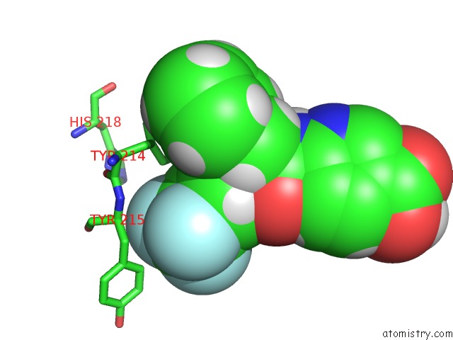
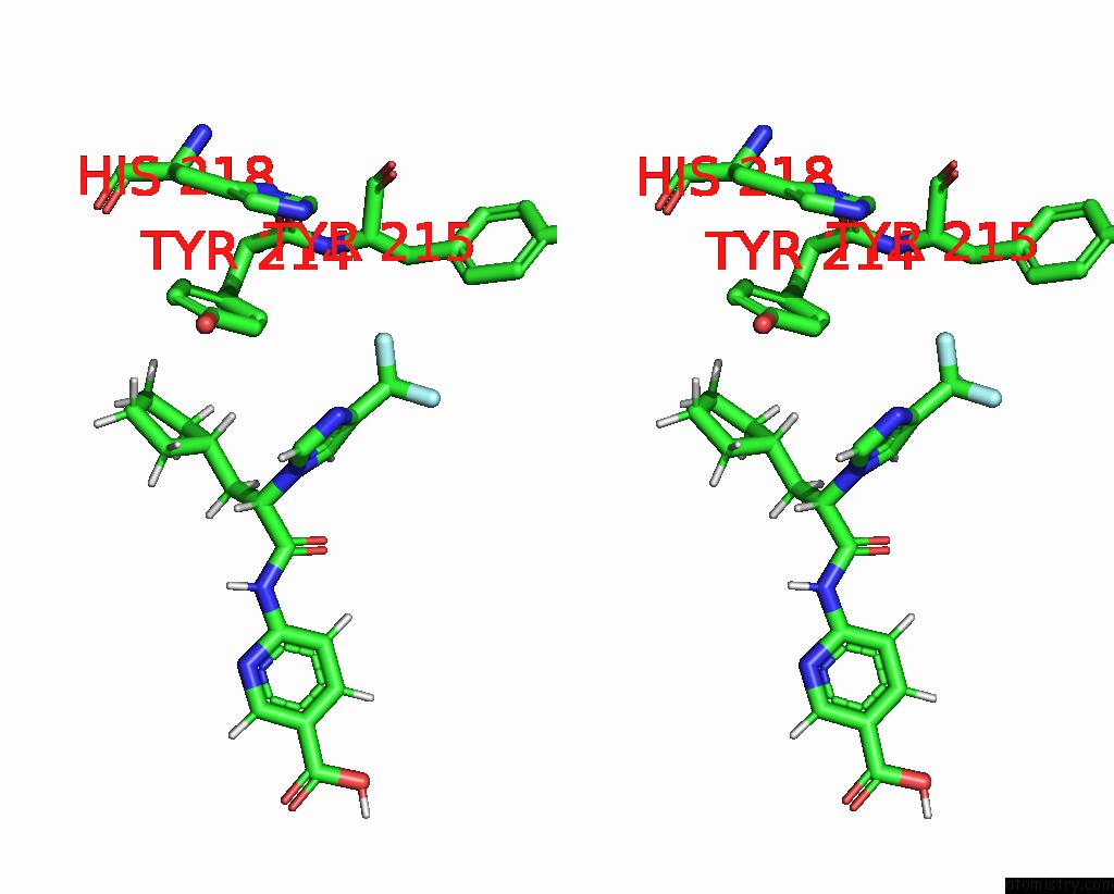
Fluorine binding site 1 out of 3 in 3vf6
Go back to
Fluorine binding site 1 out
of 3 in the Glucokinase in Complex with Glucose and Activator
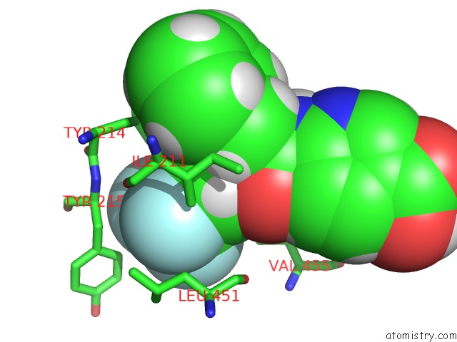
Mono view
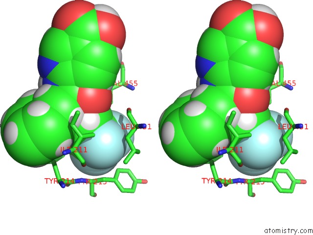
Stereo pair view
Mono view
Stereo pair view
A full contact list of Fluorine with other atoms in the F binding
site number 1 of Glucokinase in Complex with Glucose and Activator within 5.0Å range:
|
Fluorine binding site 2 out of 3 in 3vf6
Go back to
Fluorine binding site 2 out
of 3 in the Glucokinase in Complex with Glucose and Activator
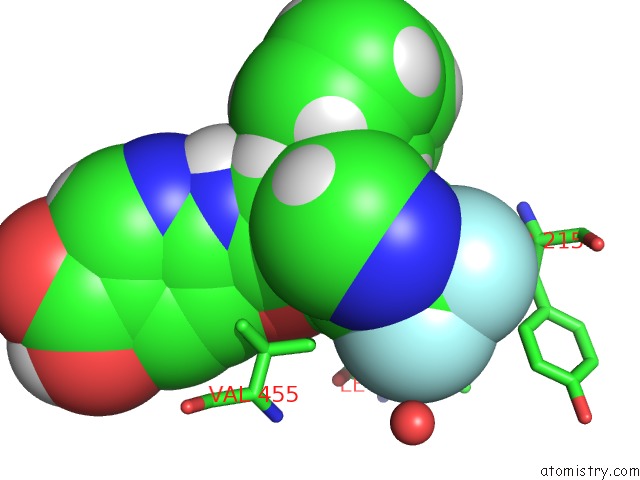
Mono view
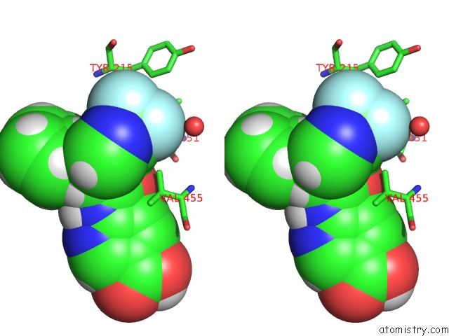
Stereo pair view
Mono view
Stereo pair view
A full contact list of Fluorine with other atoms in the F binding
site number 2 of Glucokinase in Complex with Glucose and Activator within 5.0Å range:
|
Fluorine binding site 3 out of 3 in 3vf6
Go back to
Fluorine binding site 3 out
of 3 in the Glucokinase in Complex with Glucose and Activator
Mono view
Stereo pair view
Mono view
Stereo pair view
A full contact list of Fluorine with other atoms in the F binding
site number 3 of Glucokinase in Complex with Glucose and Activator within 5.0Å range:
|
Reference:
S.Liu,
M.J.Ammirati,
X.Song,
J.D.Knafels,
J.Zhang,
S.E.Greasley,
J.A.Pfefferkorn,
X.Qiu.
Insights Into Mechanism of Glucokinase Activation: Observation of Multiple Distinct Protein Conformations. J.Biol.Chem. V. 287 13598 2012.
ISSN: ISSN 0021-9258
PubMed: 22298776
DOI: 10.1074/JBC.M111.274126
Page generated: Wed Jul 31 23:16:12 2024
ISSN: ISSN 0021-9258
PubMed: 22298776
DOI: 10.1074/JBC.M111.274126
Last articles
Cl in 7TRVCl in 7TSI
Cl in 7TSG
Cl in 7TSH
Cl in 7TRU
Cl in 7TPM
Cl in 7TRT
Cl in 7TRP
Cl in 7TQM
Cl in 7TP6