Fluorine »
PDB 3vrs-3wyk »
3vw7 »
Fluorine in PDB 3vw7: Crystal Structure of Human Protease-Activated Receptor 1 (PAR1) Bound with Antagonist Vorapaxar at 2.2 Angstrom
Enzymatic activity of Crystal Structure of Human Protease-Activated Receptor 1 (PAR1) Bound with Antagonist Vorapaxar at 2.2 Angstrom
All present enzymatic activity of Crystal Structure of Human Protease-Activated Receptor 1 (PAR1) Bound with Antagonist Vorapaxar at 2.2 Angstrom:
3.2.1.17;
3.2.1.17;
Protein crystallography data
The structure of Crystal Structure of Human Protease-Activated Receptor 1 (PAR1) Bound with Antagonist Vorapaxar at 2.2 Angstrom, PDB code: 3vw7
was solved by
C.Zhang,
Y.Srinivasan,
D.H.Arlow,
J.J.Fung,
D.Palmer,
Y.Zheng,
H.F.Green,
A.Pandey,
R.O.Dror,
D.E.Shaw,
W.I.Weis,
S.R.Coughlin,
B.K.Kobilka,
with X-Ray Crystallography technique. A brief refinement statistics is given in the table below:
| Resolution Low / High (Å) | 28.27 / 2.20 |
| Space group | P 21 21 21 |
| Cell size a, b, c (Å), α, β, γ (°) | 44.044, 71.460, 172.187, 90.00, 90.00, 90.00 |
| R / Rfree (%) | 21.8 / 23.5 |
Other elements in 3vw7:
The structure of Crystal Structure of Human Protease-Activated Receptor 1 (PAR1) Bound with Antagonist Vorapaxar at 2.2 Angstrom also contains other interesting chemical elements:
| Chlorine | (Cl) | 1 atom |
| Sodium | (Na) | 1 atom |
Fluorine Binding Sites:
The binding sites of Fluorine atom in the Crystal Structure of Human Protease-Activated Receptor 1 (PAR1) Bound with Antagonist Vorapaxar at 2.2 Angstrom
(pdb code 3vw7). This binding sites where shown within
5.0 Angstroms radius around Fluorine atom.
In total only one binding site of Fluorine was determined in the Crystal Structure of Human Protease-Activated Receptor 1 (PAR1) Bound with Antagonist Vorapaxar at 2.2 Angstrom, PDB code: 3vw7:
In total only one binding site of Fluorine was determined in the Crystal Structure of Human Protease-Activated Receptor 1 (PAR1) Bound with Antagonist Vorapaxar at 2.2 Angstrom, PDB code: 3vw7:
Fluorine binding site 1 out of 1 in 3vw7
Go back to
Fluorine binding site 1 out
of 1 in the Crystal Structure of Human Protease-Activated Receptor 1 (PAR1) Bound with Antagonist Vorapaxar at 2.2 Angstrom
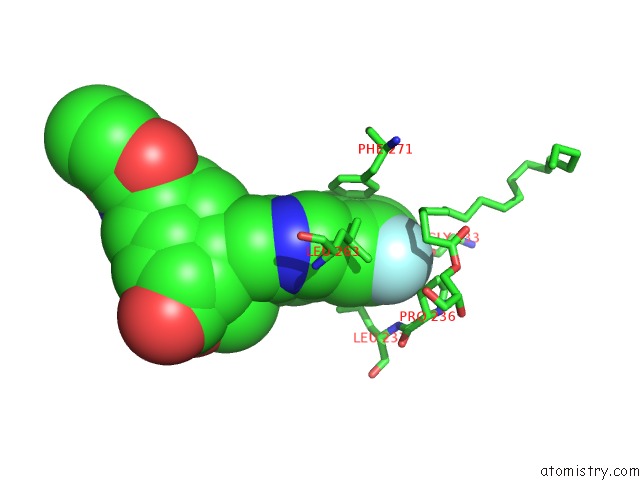
Mono view
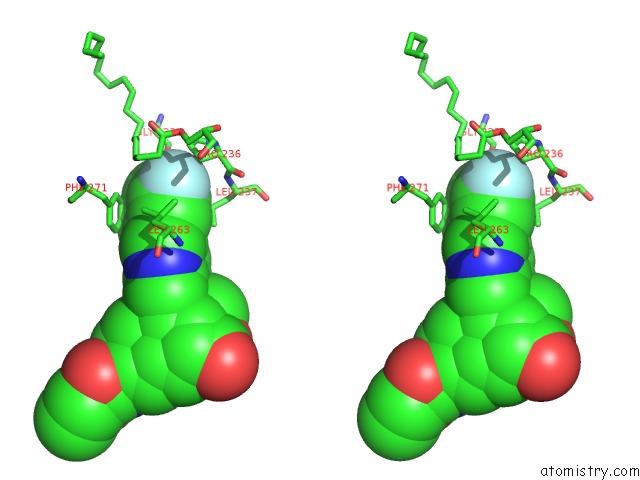
Stereo pair view
Mono view
Stereo pair view
A full contact list of Fluorine with other atoms in the F binding
site number 1 of Crystal Structure of Human Protease-Activated Receptor 1 (PAR1) Bound with Antagonist Vorapaxar at 2.2 Angstrom within 5.0Å range:
|
Reference:
C.Zhang,
Y.Srinivasan,
D.H.Arlow,
J.J.Fung,
D.Palmer,
Y.Zheng,
H.F.Green,
A.Pandey,
R.O.Dror,
D.E.Shaw,
W.I.Weis,
S.R.Coughlin,
B.K.Kobilka.
High-Resolution Crystal Structure of Human Protease-Activated Receptor 1 Nature V. 492 387 2012.
ISSN: ISSN 0028-0836
PubMed: 23222541
DOI: 10.1038/NATURE11701
Page generated: Mon Jul 14 19:57:48 2025
ISSN: ISSN 0028-0836
PubMed: 23222541
DOI: 10.1038/NATURE11701
Last articles
F in 4PYYF in 4PZH
F in 4PYX
F in 4PX5
F in 4PY4
F in 4PWL
F in 4PVU
F in 4PUW
F in 4PU9
F in 4PUU