Fluorine »
PDB 3vrs-3wyk »
3wsp »
Fluorine in PDB 3wsp: Crystal Structure of P450BM3 with N-Perfluorononanoyl-L-Tryptophan
Enzymatic activity of Crystal Structure of P450BM3 with N-Perfluorononanoyl-L-Tryptophan
All present enzymatic activity of Crystal Structure of P450BM3 with N-Perfluorononanoyl-L-Tryptophan:
1.14.14.1; 1.6.2.4;
1.14.14.1; 1.6.2.4;
Protein crystallography data
The structure of Crystal Structure of P450BM3 with N-Perfluorononanoyl-L-Tryptophan, PDB code: 3wsp
was solved by
Z.Cong,
O.Shoji,
C.Kasai,
H.Sugimoto,
Y.Shiro,
Y.Watanabe,
with X-Ray Crystallography technique. A brief refinement statistics is given in the table below:
| Resolution Low / High (Å) | 19.96 / 1.80 |
| Space group | P 1 21 1 |
| Cell size a, b, c (Å), α, β, γ (°) | 58.875, 145.392, 63.029, 90.00, 97.06, 90.00 |
| R / Rfree (%) | 17.4 / 21.2 |
Other elements in 3wsp:
The structure of Crystal Structure of P450BM3 with N-Perfluorononanoyl-L-Tryptophan also contains other interesting chemical elements:
| Iron | (Fe) | 2 atoms |
Fluorine Binding Sites:
Pages:
>>> Page 1 <<< Page 2, Binding sites: 11 - 20; Page 3, Binding sites: 21 - 30; Page 4, Binding sites: 31 - 34;Binding sites:
The binding sites of Fluorine atom in the Crystal Structure of P450BM3 with N-Perfluorononanoyl-L-Tryptophan (pdb code 3wsp). This binding sites where shown within 5.0 Angstroms radius around Fluorine atom.In total 34 binding sites of Fluorine where determined in the Crystal Structure of P450BM3 with N-Perfluorononanoyl-L-Tryptophan, PDB code: 3wsp:
Jump to Fluorine binding site number: 1; 2; 3; 4; 5; 6; 7; 8; 9; 10;
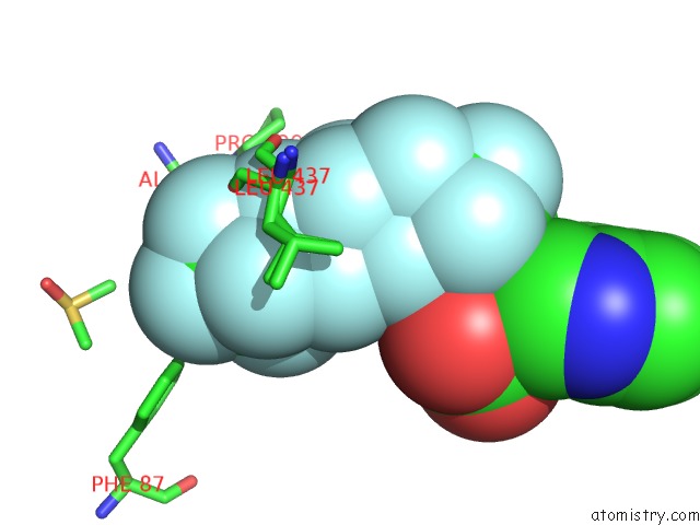
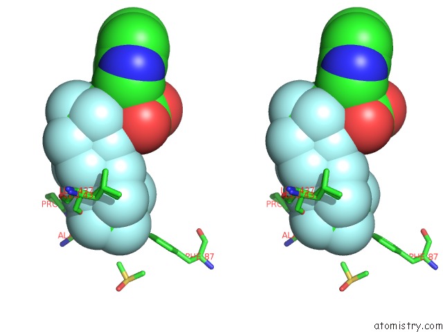
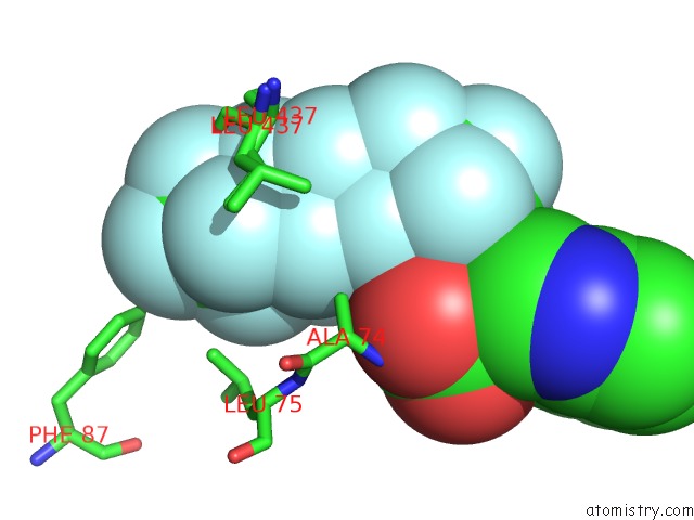
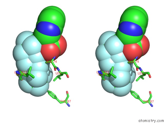
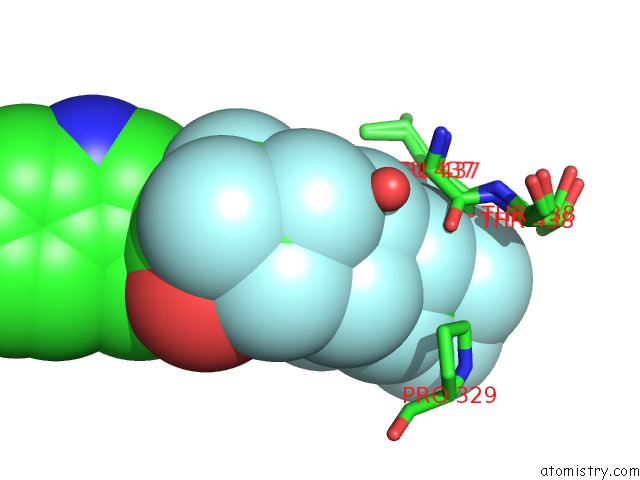
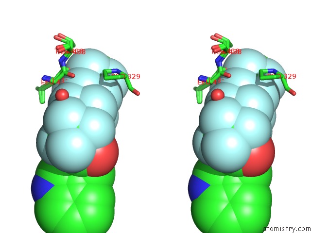
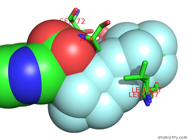
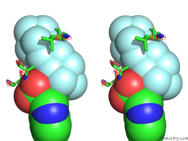
Fluorine binding site 1 out of 34 in 3wsp
Go back to
Fluorine binding site 1 out
of 34 in the Crystal Structure of P450BM3 with N-Perfluorononanoyl-L-Tryptophan
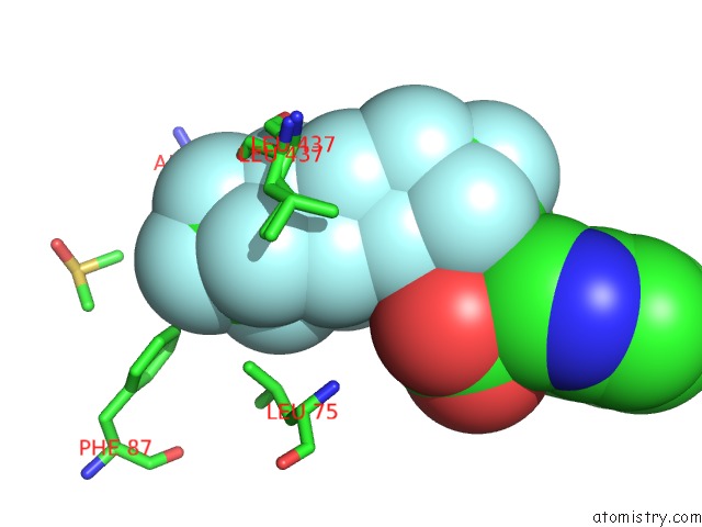
Mono view
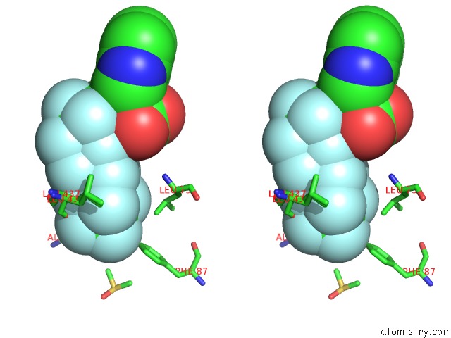
Stereo pair view
Mono view
Stereo pair view
A full contact list of Fluorine with other atoms in the F binding
site number 1 of Crystal Structure of P450BM3 with N-Perfluorononanoyl-L-Tryptophan within 5.0Å range:
|
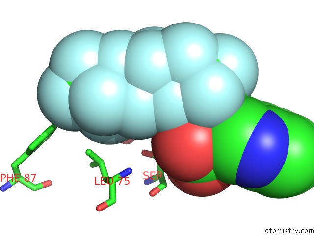
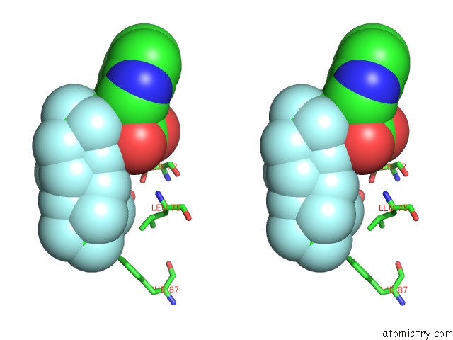
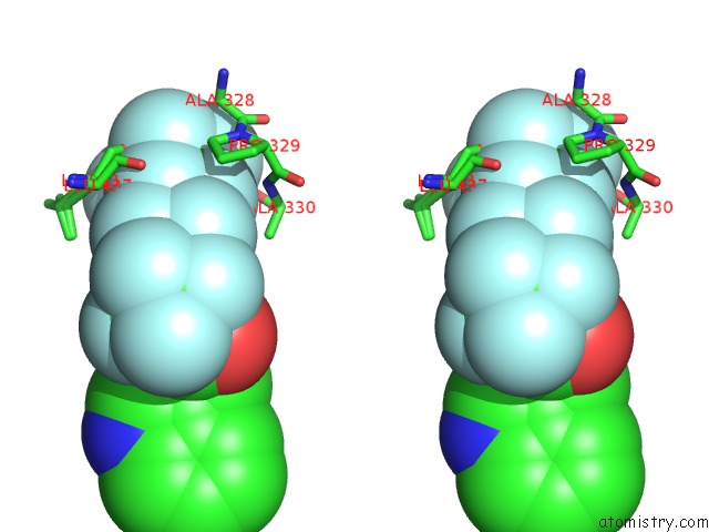
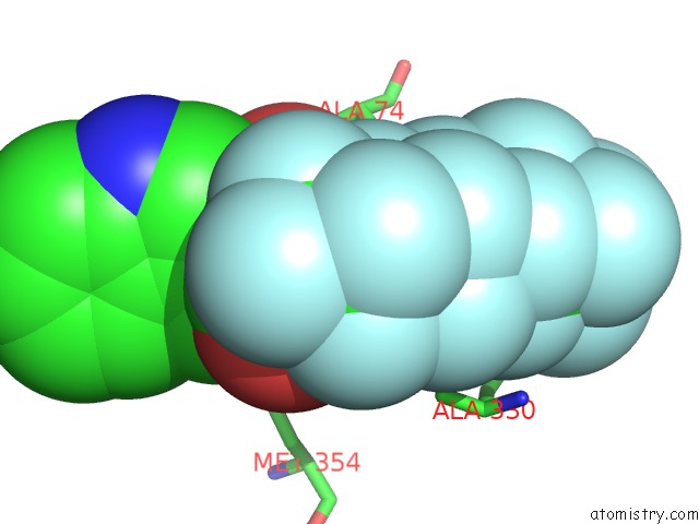
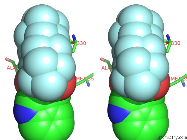
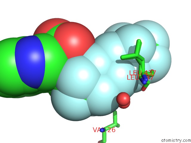
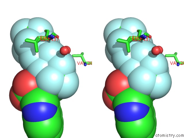
Fluorine binding site 2 out of 34 in 3wsp
Go back to
Fluorine binding site 2 out
of 34 in the Crystal Structure of P450BM3 with N-Perfluorononanoyl-L-Tryptophan
Mono view
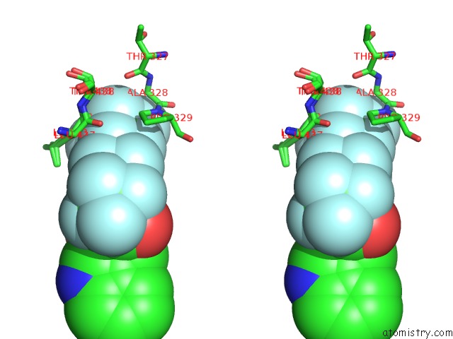
Stereo pair view
Mono view
Stereo pair view
A full contact list of Fluorine with other atoms in the F binding
site number 2 of Crystal Structure of P450BM3 with N-Perfluorononanoyl-L-Tryptophan within 5.0Å range:
|
Fluorine binding site 3 out of 34 in 3wsp
Go back to
Fluorine binding site 3 out
of 34 in the Crystal Structure of P450BM3 with N-Perfluorononanoyl-L-Tryptophan
Mono view
Stereo pair view
Mono view
Stereo pair view
A full contact list of Fluorine with other atoms in the F binding
site number 3 of Crystal Structure of P450BM3 with N-Perfluorononanoyl-L-Tryptophan within 5.0Å range:
|
Fluorine binding site 4 out of 34 in 3wsp
Go back to
Fluorine binding site 4 out
of 34 in the Crystal Structure of P450BM3 with N-Perfluorononanoyl-L-Tryptophan
Mono view
Stereo pair view
Mono view
Stereo pair view
A full contact list of Fluorine with other atoms in the F binding
site number 4 of Crystal Structure of P450BM3 with N-Perfluorononanoyl-L-Tryptophan within 5.0Å range:
|
Fluorine binding site 5 out of 34 in 3wsp
Go back to
Fluorine binding site 5 out
of 34 in the Crystal Structure of P450BM3 with N-Perfluorononanoyl-L-Tryptophan
Mono view
Stereo pair view
Mono view
Stereo pair view
A full contact list of Fluorine with other atoms in the F binding
site number 5 of Crystal Structure of P450BM3 with N-Perfluorononanoyl-L-Tryptophan within 5.0Å range:
|
Fluorine binding site 6 out of 34 in 3wsp
Go back to
Fluorine binding site 6 out
of 34 in the Crystal Structure of P450BM3 with N-Perfluorononanoyl-L-Tryptophan
Mono view
Stereo pair view
Mono view
Stereo pair view
A full contact list of Fluorine with other atoms in the F binding
site number 6 of Crystal Structure of P450BM3 with N-Perfluorononanoyl-L-Tryptophan within 5.0Å range:
|
Fluorine binding site 7 out of 34 in 3wsp
Go back to
Fluorine binding site 7 out
of 34 in the Crystal Structure of P450BM3 with N-Perfluorononanoyl-L-Tryptophan
Mono view
Stereo pair view
Mono view
Stereo pair view
A full contact list of Fluorine with other atoms in the F binding
site number 7 of Crystal Structure of P450BM3 with N-Perfluorononanoyl-L-Tryptophan within 5.0Å range:
|
Fluorine binding site 8 out of 34 in 3wsp
Go back to
Fluorine binding site 8 out
of 34 in the Crystal Structure of P450BM3 with N-Perfluorononanoyl-L-Tryptophan
Mono view
Stereo pair view
Mono view
Stereo pair view
A full contact list of Fluorine with other atoms in the F binding
site number 8 of Crystal Structure of P450BM3 with N-Perfluorononanoyl-L-Tryptophan within 5.0Å range:
|
Fluorine binding site 9 out of 34 in 3wsp
Go back to
Fluorine binding site 9 out
of 34 in the Crystal Structure of P450BM3 with N-Perfluorononanoyl-L-Tryptophan
Mono view
Stereo pair view
Mono view
Stereo pair view
A full contact list of Fluorine with other atoms in the F binding
site number 9 of Crystal Structure of P450BM3 with N-Perfluorononanoyl-L-Tryptophan within 5.0Å range:
|
Fluorine binding site 10 out of 34 in 3wsp
Go back to
Fluorine binding site 10 out
of 34 in the Crystal Structure of P450BM3 with N-Perfluorononanoyl-L-Tryptophan
Mono view
Stereo pair view
Mono view
Stereo pair view
A full contact list of Fluorine with other atoms in the F binding
site number 10 of Crystal Structure of P450BM3 with N-Perfluorononanoyl-L-Tryptophan within 5.0Å range:
|
Reference:
Z.Cong,
O.Shoji,
C.Kasai,
N.Kawakami,
H.Sugimoto,
Y.Shiro,
Y.Watanabe.
Activation of Wild-Type Cytochrome P450BM3 By the Next Generation of Decoy Molecules: Enhanced Hydroxylation of Gaseous Alkanes and Crystallographic Evidence. Acs Catalysis.
ISSN: ESSN 2155-5435
DOI: 10.1021/CS501592F
Page generated: Mon Jul 14 20:06:49 2025
ISSN: ESSN 2155-5435
DOI: 10.1021/CS501592F
Last articles
Fe in 2YXOFe in 2YRS
Fe in 2YXC
Fe in 2YNM
Fe in 2YVJ
Fe in 2YP1
Fe in 2YU2
Fe in 2YU1
Fe in 2YQB
Fe in 2YOO