Fluorine »
PDB 3wyl-460d »
3wze »
Fluorine in PDB 3wze: Kdr in Complex with Ligand Sorafenib
Enzymatic activity of Kdr in Complex with Ligand Sorafenib
All present enzymatic activity of Kdr in Complex with Ligand Sorafenib:
2.7.10.1;
2.7.10.1;
Protein crystallography data
The structure of Kdr in Complex with Ligand Sorafenib, PDB code: 3wze
was solved by
K.Okamoto,
M.Ikemori_Kawada,
A.Inoue,
J.Matsui,
with X-Ray Crystallography technique. A brief refinement statistics is given in the table below:
| Resolution Low / High (Å) | 67.78 / 1.90 |
| Space group | C 1 2 1 |
| Cell size a, b, c (Å), α, β, γ (°) | 135.921, 57.357, 52.170, 90.00, 94.17, 90.00 |
| R / Rfree (%) | 18.5 / 22.4 |
Other elements in 3wze:
The structure of Kdr in Complex with Ligand Sorafenib also contains other interesting chemical elements:
| Chlorine | (Cl) | 1 atom |
Fluorine Binding Sites:
The binding sites of Fluorine atom in the Kdr in Complex with Ligand Sorafenib
(pdb code 3wze). This binding sites where shown within
5.0 Angstroms radius around Fluorine atom.
In total 3 binding sites of Fluorine where determined in the Kdr in Complex with Ligand Sorafenib, PDB code: 3wze:
Jump to Fluorine binding site number: 1; 2; 3;
In total 3 binding sites of Fluorine where determined in the Kdr in Complex with Ligand Sorafenib, PDB code: 3wze:
Jump to Fluorine binding site number: 1; 2; 3;
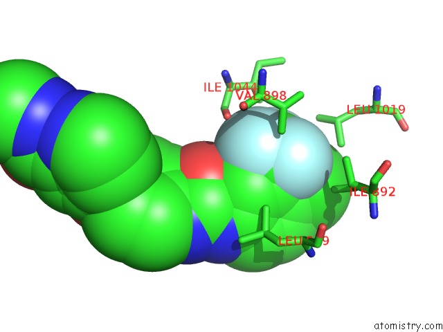
Fluorine binding site 1 out of 3 in 3wze
Go back to
Fluorine binding site 1 out
of 3 in the Kdr in Complex with Ligand Sorafenib

Mono view

Stereo pair view
Mono view
Stereo pair view
A full contact list of Fluorine with other atoms in the F binding
site number 1 of Kdr in Complex with Ligand Sorafenib within 5.0Å range:
|
Fluorine binding site 2 out of 3 in 3wze
Go back to
Fluorine binding site 2 out
of 3 in the Kdr in Complex with Ligand Sorafenib

Mono view

Stereo pair view
Mono view
Stereo pair view
A full contact list of Fluorine with other atoms in the F binding
site number 2 of Kdr in Complex with Ligand Sorafenib within 5.0Å range:
|
Fluorine binding site 3 out of 3 in 3wze
Go back to
Fluorine binding site 3 out
of 3 in the Kdr in Complex with Ligand Sorafenib
Mono view

Stereo pair view
Mono view
Stereo pair view
A full contact list of Fluorine with other atoms in the F binding
site number 3 of Kdr in Complex with Ligand Sorafenib within 5.0Å range:
|
Reference:
K.Okamoto,
M.Ikemori-Kawada,
A.Jestel,
K.Von Konig,
Y.Funahashi,
T.Matsushima,
A.Tsuruoka,
A.Inoue,
J.Matsui.
Distinct Binding Mode of Multikinase Inhibitor Lenvatinib Revealed By Biochemical Characterization. Acs Med.Chem.Lett. V. 6 89 2015.
ISSN: ISSN 1948-5875
PubMed: 25589937
DOI: 10.1021/ML500394M
Page generated: Wed Jul 31 23:41:27 2024
ISSN: ISSN 1948-5875
PubMed: 25589937
DOI: 10.1021/ML500394M
Last articles
Zn in 9J0NZn in 9J0O
Zn in 9J0P
Zn in 9FJX
Zn in 9EKB
Zn in 9C0F
Zn in 9CAH
Zn in 9CH0
Zn in 9CH3
Zn in 9CH1