Fluorine »
PDB 3wyl-460d »
3zse »
Fluorine in PDB 3zse: 3D Structure of A Thermophilic Family GH11 Xylanase From Thermobifida Fusca
Enzymatic activity of 3D Structure of A Thermophilic Family GH11 Xylanase From Thermobifida Fusca
All present enzymatic activity of 3D Structure of A Thermophilic Family GH11 Xylanase From Thermobifida Fusca:
3.2.1.8;
3.2.1.8;
Protein crystallography data
The structure of 3D Structure of A Thermophilic Family GH11 Xylanase From Thermobifida Fusca, PDB code: 3zse
was solved by
A.Lammerts Van Bueren,
S.Otani,
E.P.Friis,
K.S Wilson,
G.J.Davies,
with X-Ray Crystallography technique. A brief refinement statistics is given in the table below:
| Resolution Low / High (Å) | 37.29 / 1.78 |
| Space group | P 21 21 21 |
| Cell size a, b, c (Å), α, β, γ (°) | 40.630, 41.310, 93.970, 90.00, 90.00, 90.00 |
| R / Rfree (%) | 19.34 / 24.107 |
Fluorine Binding Sites:
The binding sites of Fluorine atom in the 3D Structure of A Thermophilic Family GH11 Xylanase From Thermobifida Fusca
(pdb code 3zse). This binding sites where shown within
5.0 Angstroms radius around Fluorine atom.
In total only one binding site of Fluorine was determined in the 3D Structure of A Thermophilic Family GH11 Xylanase From Thermobifida Fusca, PDB code: 3zse:
In total only one binding site of Fluorine was determined in the 3D Structure of A Thermophilic Family GH11 Xylanase From Thermobifida Fusca, PDB code: 3zse:
Fluorine binding site 1 out of 1 in 3zse
Go back to
Fluorine binding site 1 out
of 1 in the 3D Structure of A Thermophilic Family GH11 Xylanase From Thermobifida Fusca
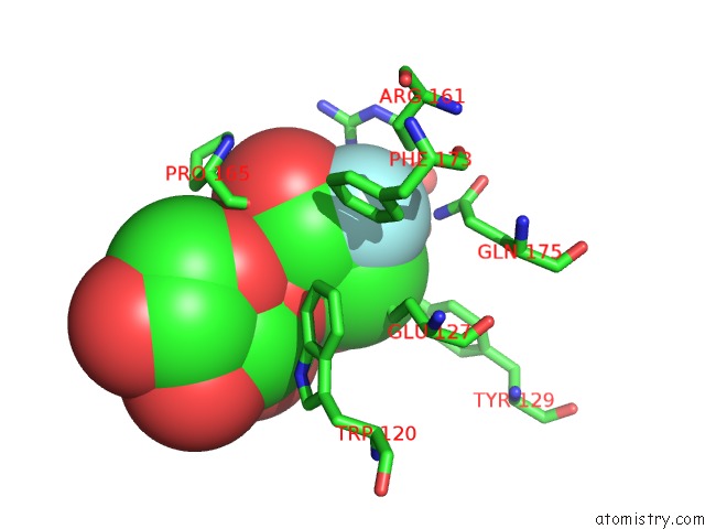
Mono view
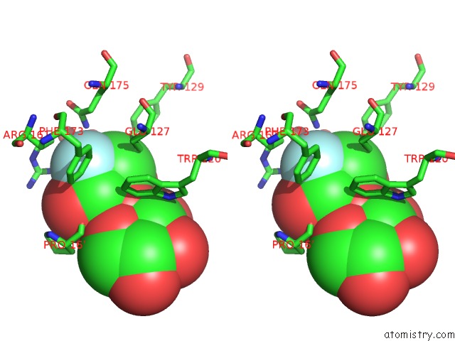
Stereo pair view
Mono view
Stereo pair view
A full contact list of Fluorine with other atoms in the F binding
site number 1 of 3D Structure of A Thermophilic Family GH11 Xylanase From Thermobifida Fusca within 5.0Å range:
|
Reference:
A.Lammerts Van Bueren,
S.Otani,
E.P.Friis,
K.S.Wilson,
G.J.Davies.
Three-Dimensional Structure of A Thermophilic Family GH11 Xylanase From Thermobifida Fusca. Acta Crystallogr.,Sect.F V. 68 141 2012.
ISSN: ISSN 1744-3091
PubMed: 22297985
DOI: 10.1107/S1744309111049608
Page generated: Wed Jul 31 23:49:58 2024
ISSN: ISSN 1744-3091
PubMed: 22297985
DOI: 10.1107/S1744309111049608
Last articles
Zn in 9J0NZn in 9J0O
Zn in 9J0P
Zn in 9FJX
Zn in 9EKB
Zn in 9C0F
Zn in 9CAH
Zn in 9CH0
Zn in 9CH3
Zn in 9CH1