Fluorine »
PDB 3wyl-460d »
3zwv »
Fluorine in PDB 3zwv: Crystal Structure of Adp-Ribosyl Cyclase Complexed with Ara-2'F-Adp- Ribose at 2.3 Angstrom
Enzymatic activity of Crystal Structure of Adp-Ribosyl Cyclase Complexed with Ara-2'F-Adp- Ribose at 2.3 Angstrom
All present enzymatic activity of Crystal Structure of Adp-Ribosyl Cyclase Complexed with Ara-2'F-Adp- Ribose at 2.3 Angstrom:
3.2.2.5;
3.2.2.5;
Protein crystallography data
The structure of Crystal Structure of Adp-Ribosyl Cyclase Complexed with Ara-2'F-Adp- Ribose at 2.3 Angstrom, PDB code: 3zwv
was solved by
M.Kotaka,
R.Graeff,
L.H.Zhang,
H.C.Lee,
Q.Hao,
with X-Ray Crystallography technique. A brief refinement statistics is given in the table below:
| Resolution Low / High (Å) | 30.00 / 2.30 |
| Space group | P 1 |
| Cell size a, b, c (Å), α, β, γ (°) | 61.146, 76.481, 141.366, 88.09, 90.88, 91.02 |
| R / Rfree (%) | 21.5 / 27.7 |
Fluorine Binding Sites:
The binding sites of Fluorine atom in the Crystal Structure of Adp-Ribosyl Cyclase Complexed with Ara-2'F-Adp- Ribose at 2.3 Angstrom
(pdb code 3zwv). This binding sites where shown within
5.0 Angstroms radius around Fluorine atom.
In total 8 binding sites of Fluorine where determined in the Crystal Structure of Adp-Ribosyl Cyclase Complexed with Ara-2'F-Adp- Ribose at 2.3 Angstrom, PDB code: 3zwv:
Jump to Fluorine binding site number: 1; 2; 3; 4; 5; 6; 7; 8;
In total 8 binding sites of Fluorine where determined in the Crystal Structure of Adp-Ribosyl Cyclase Complexed with Ara-2'F-Adp- Ribose at 2.3 Angstrom, PDB code: 3zwv:
Jump to Fluorine binding site number: 1; 2; 3; 4; 5; 6; 7; 8;
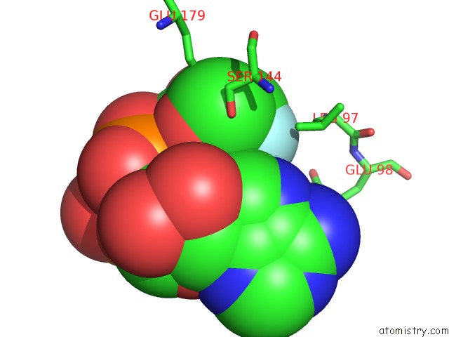
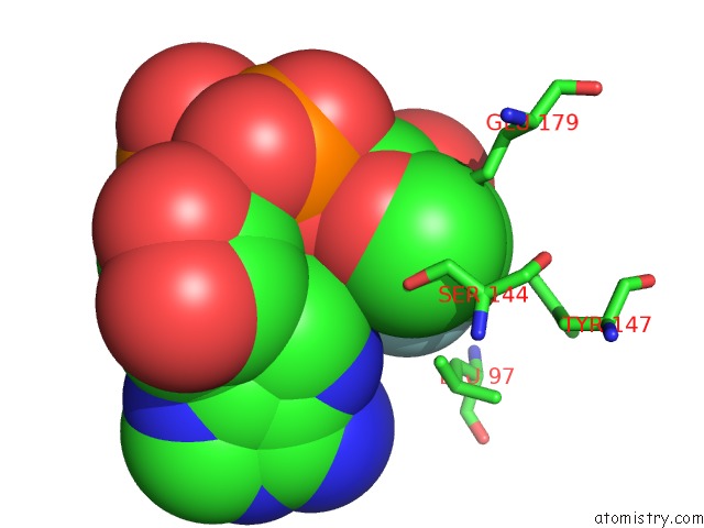
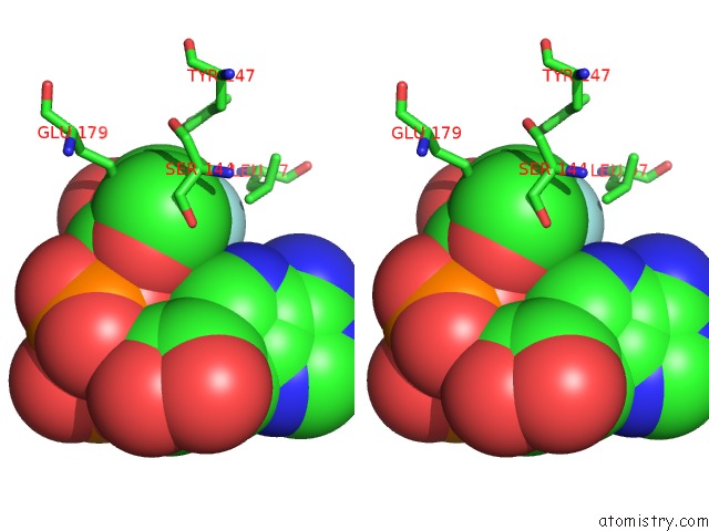
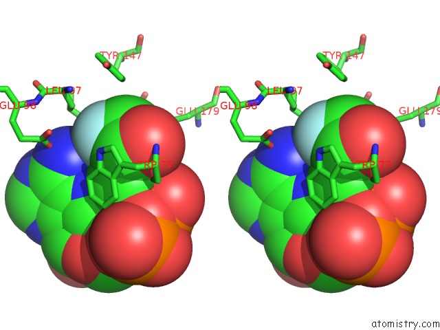
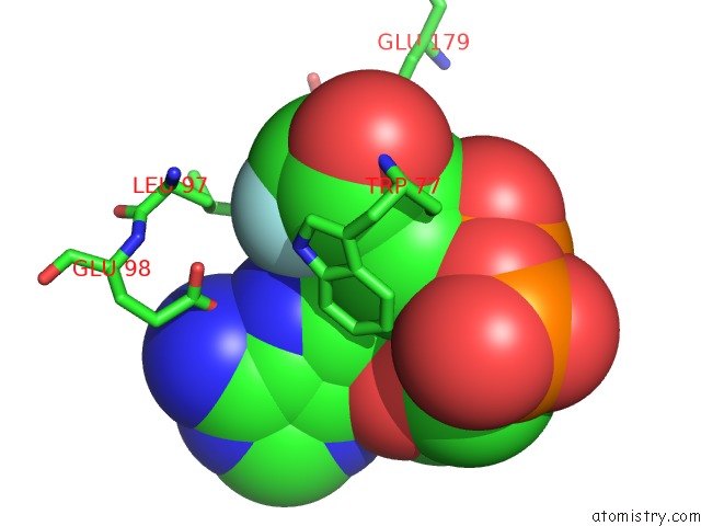
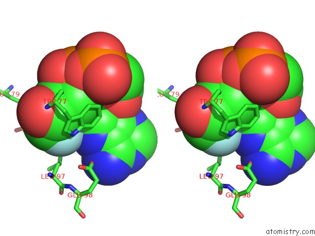
Fluorine binding site 1 out of 8 in 3zwv
Go back to
Fluorine binding site 1 out
of 8 in the Crystal Structure of Adp-Ribosyl Cyclase Complexed with Ara-2'F-Adp- Ribose at 2.3 Angstrom
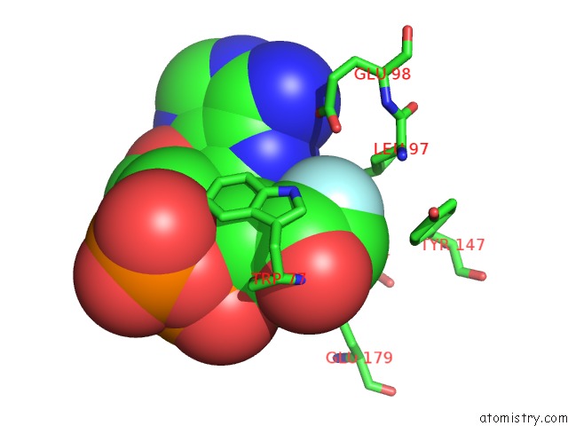
Mono view
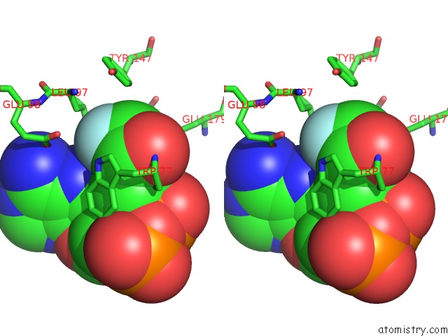
Stereo pair view
Mono view
Stereo pair view
A full contact list of Fluorine with other atoms in the F binding
site number 1 of Crystal Structure of Adp-Ribosyl Cyclase Complexed with Ara-2'F-Adp- Ribose at 2.3 Angstrom within 5.0Å range:
|
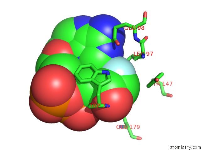
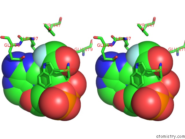
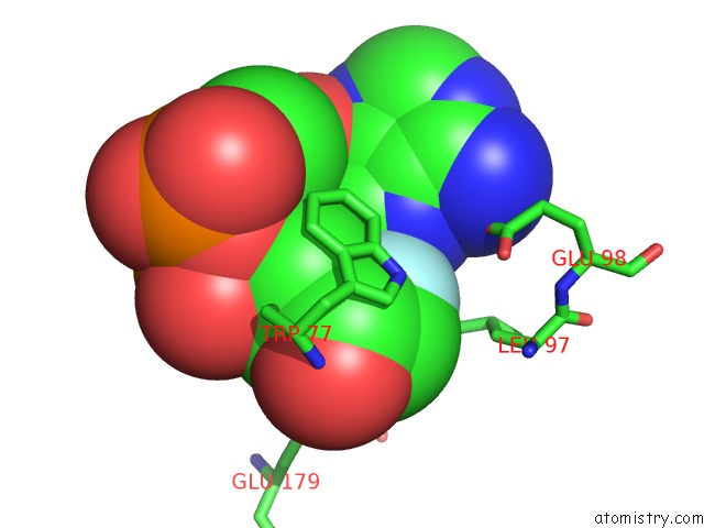
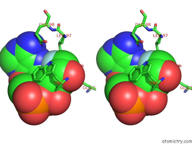
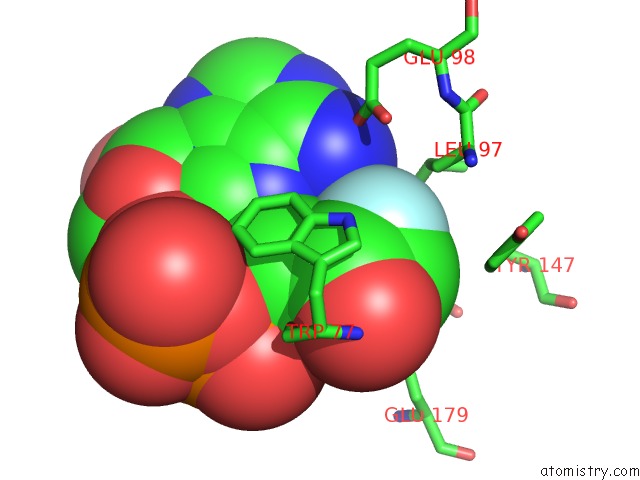
Fluorine binding site 2 out of 8 in 3zwv
Go back to
Fluorine binding site 2 out
of 8 in the Crystal Structure of Adp-Ribosyl Cyclase Complexed with Ara-2'F-Adp- Ribose at 2.3 Angstrom
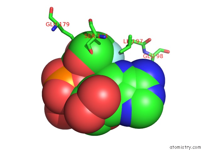
Mono view
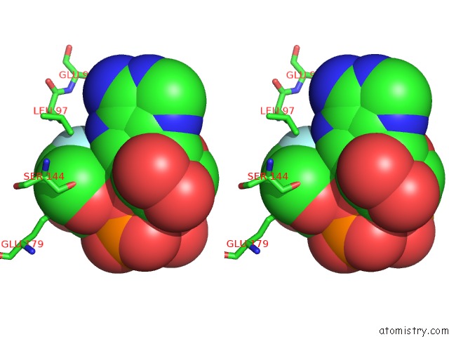
Stereo pair view
Mono view
Stereo pair view
A full contact list of Fluorine with other atoms in the F binding
site number 2 of Crystal Structure of Adp-Ribosyl Cyclase Complexed with Ara-2'F-Adp- Ribose at 2.3 Angstrom within 5.0Å range:
|
Fluorine binding site 3 out of 8 in 3zwv
Go back to
Fluorine binding site 3 out
of 8 in the Crystal Structure of Adp-Ribosyl Cyclase Complexed with Ara-2'F-Adp- Ribose at 2.3 Angstrom
Mono view

Stereo pair view
Mono view
Stereo pair view
A full contact list of Fluorine with other atoms in the F binding
site number 3 of Crystal Structure of Adp-Ribosyl Cyclase Complexed with Ara-2'F-Adp- Ribose at 2.3 Angstrom within 5.0Å range:
|
Fluorine binding site 4 out of 8 in 3zwv
Go back to
Fluorine binding site 4 out
of 8 in the Crystal Structure of Adp-Ribosyl Cyclase Complexed with Ara-2'F-Adp- Ribose at 2.3 Angstrom
Mono view
Stereo pair view
Mono view
Stereo pair view
A full contact list of Fluorine with other atoms in the F binding
site number 4 of Crystal Structure of Adp-Ribosyl Cyclase Complexed with Ara-2'F-Adp- Ribose at 2.3 Angstrom within 5.0Å range:
|
Fluorine binding site 5 out of 8 in 3zwv
Go back to
Fluorine binding site 5 out
of 8 in the Crystal Structure of Adp-Ribosyl Cyclase Complexed with Ara-2'F-Adp- Ribose at 2.3 Angstrom
Mono view
Stereo pair view
Mono view
Stereo pair view
A full contact list of Fluorine with other atoms in the F binding
site number 5 of Crystal Structure of Adp-Ribosyl Cyclase Complexed with Ara-2'F-Adp- Ribose at 2.3 Angstrom within 5.0Å range:
|
Fluorine binding site 6 out of 8 in 3zwv
Go back to
Fluorine binding site 6 out
of 8 in the Crystal Structure of Adp-Ribosyl Cyclase Complexed with Ara-2'F-Adp- Ribose at 2.3 Angstrom
Mono view
Stereo pair view
Mono view
Stereo pair view
A full contact list of Fluorine with other atoms in the F binding
site number 6 of Crystal Structure of Adp-Ribosyl Cyclase Complexed with Ara-2'F-Adp- Ribose at 2.3 Angstrom within 5.0Å range:
|
Fluorine binding site 7 out of 8 in 3zwv
Go back to
Fluorine binding site 7 out
of 8 in the Crystal Structure of Adp-Ribosyl Cyclase Complexed with Ara-2'F-Adp- Ribose at 2.3 Angstrom
Mono view
Stereo pair view
Mono view
Stereo pair view
A full contact list of Fluorine with other atoms in the F binding
site number 7 of Crystal Structure of Adp-Ribosyl Cyclase Complexed with Ara-2'F-Adp- Ribose at 2.3 Angstrom within 5.0Å range:
|
Fluorine binding site 8 out of 8 in 3zwv
Go back to
Fluorine binding site 8 out
of 8 in the Crystal Structure of Adp-Ribosyl Cyclase Complexed with Ara-2'F-Adp- Ribose at 2.3 Angstrom
Mono view
Stereo pair view
Mono view
Stereo pair view
A full contact list of Fluorine with other atoms in the F binding
site number 8 of Crystal Structure of Adp-Ribosyl Cyclase Complexed with Ara-2'F-Adp- Ribose at 2.3 Angstrom within 5.0Å range:
|
Reference:
M.Kotaka,
R.Graeff,
Z.Chen,
L.H.Zhang,
H.C.Lee,
Q.Hao.
Structural Studies of Intermediates Along the Cyclization Pathway of Aplysia Adp-Ribosyl Cyclase. J. Mol. Biol. V. 415 514 2012.
ISSN: ESSN 1089-8638
PubMed: 22138343
DOI: 10.1016/J.JMB.2011.11.022
Page generated: Wed Jul 31 23:50:57 2024
ISSN: ESSN 1089-8638
PubMed: 22138343
DOI: 10.1016/J.JMB.2011.11.022
Last articles
Zn in 9J0NZn in 9J0O
Zn in 9J0P
Zn in 9FJX
Zn in 9EKB
Zn in 9C0F
Zn in 9CAH
Zn in 9CH0
Zn in 9CH3
Zn in 9CH1