Fluorine »
PDB 4bkz-4c62 »
4c4s »
Fluorine in PDB 4c4s: Structure of Beta-Phosphoglucomutase in Complex with An Alpha-Fluorophosphonate Analogue of Beta-Glucose-1-Phosphate and Magnesium Trifluoride
Enzymatic activity of Structure of Beta-Phosphoglucomutase in Complex with An Alpha-Fluorophosphonate Analogue of Beta-Glucose-1-Phosphate and Magnesium Trifluoride
All present enzymatic activity of Structure of Beta-Phosphoglucomutase in Complex with An Alpha-Fluorophosphonate Analogue of Beta-Glucose-1-Phosphate and Magnesium Trifluoride:
5.4.2.6;
5.4.2.6;
Protein crystallography data
The structure of Structure of Beta-Phosphoglucomutase in Complex with An Alpha-Fluorophosphonate Analogue of Beta-Glucose-1-Phosphate and Magnesium Trifluoride, PDB code: 4c4s
was solved by
E.Pellegrini,
M.W.Bowler,
with X-Ray Crystallography technique. A brief refinement statistics is given in the table below:
| Resolution Low / High (Å) | 37.634 / 1.50 |
| Space group | P 21 21 21 |
| Cell size a, b, c (Å), α, β, γ (°) | 37.541, 54.337, 104.346, 90.00, 90.00, 90.00 |
| R / Rfree (%) | 17.25 / 19.84 |
Other elements in 4c4s:
The structure of Structure of Beta-Phosphoglucomutase in Complex with An Alpha-Fluorophosphonate Analogue of Beta-Glucose-1-Phosphate and Magnesium Trifluoride also contains other interesting chemical elements:
| Magnesium | (Mg) | 2 atoms |
Fluorine Binding Sites:
The binding sites of Fluorine atom in the Structure of Beta-Phosphoglucomutase in Complex with An Alpha-Fluorophosphonate Analogue of Beta-Glucose-1-Phosphate and Magnesium Trifluoride
(pdb code 4c4s). This binding sites where shown within
5.0 Angstroms radius around Fluorine atom.
In total 4 binding sites of Fluorine where determined in the Structure of Beta-Phosphoglucomutase in Complex with An Alpha-Fluorophosphonate Analogue of Beta-Glucose-1-Phosphate and Magnesium Trifluoride, PDB code: 4c4s:
Jump to Fluorine binding site number: 1; 2; 3; 4;
In total 4 binding sites of Fluorine where determined in the Structure of Beta-Phosphoglucomutase in Complex with An Alpha-Fluorophosphonate Analogue of Beta-Glucose-1-Phosphate and Magnesium Trifluoride, PDB code: 4c4s:
Jump to Fluorine binding site number: 1; 2; 3; 4;
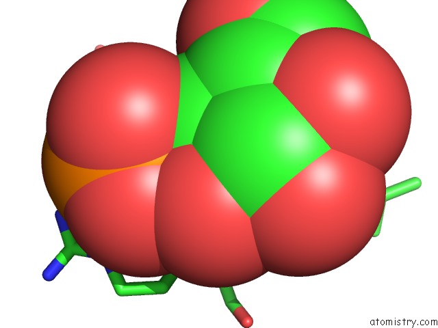
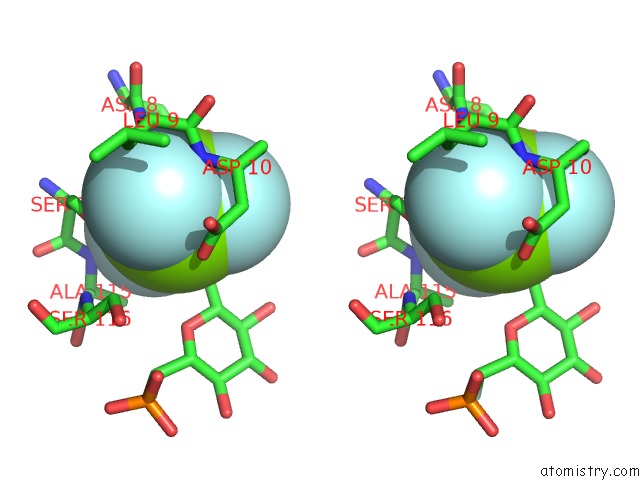
Fluorine binding site 1 out of 4 in 4c4s
Go back to
Fluorine binding site 1 out
of 4 in the Structure of Beta-Phosphoglucomutase in Complex with An Alpha-Fluorophosphonate Analogue of Beta-Glucose-1-Phosphate and Magnesium Trifluoride
Mono view

Stereo pair view
Mono view
Stereo pair view
A full contact list of Fluorine with other atoms in the F binding
site number 1 of Structure of Beta-Phosphoglucomutase in Complex with An Alpha-Fluorophosphonate Analogue of Beta-Glucose-1-Phosphate and Magnesium Trifluoride within 5.0Å range:
|
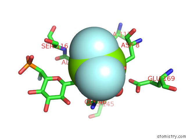
Fluorine binding site 2 out of 4 in 4c4s
Go back to
Fluorine binding site 2 out
of 4 in the Structure of Beta-Phosphoglucomutase in Complex with An Alpha-Fluorophosphonate Analogue of Beta-Glucose-1-Phosphate and Magnesium Trifluoride
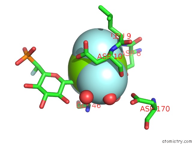
Mono view
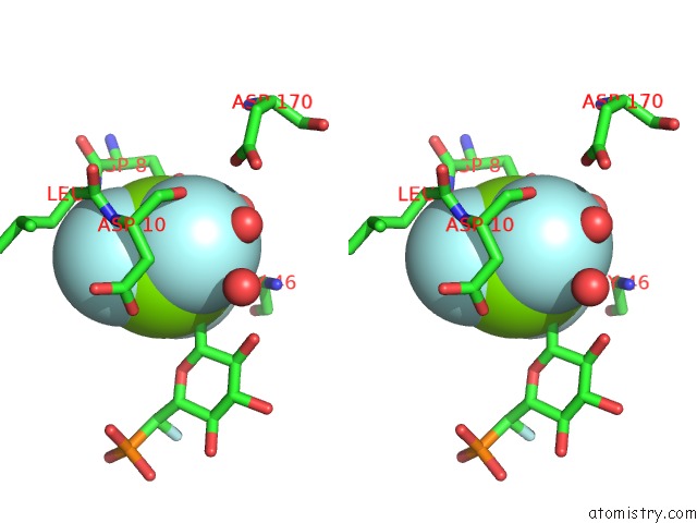
Stereo pair view
Mono view
Stereo pair view
A full contact list of Fluorine with other atoms in the F binding
site number 2 of Structure of Beta-Phosphoglucomutase in Complex with An Alpha-Fluorophosphonate Analogue of Beta-Glucose-1-Phosphate and Magnesium Trifluoride within 5.0Å range:
|
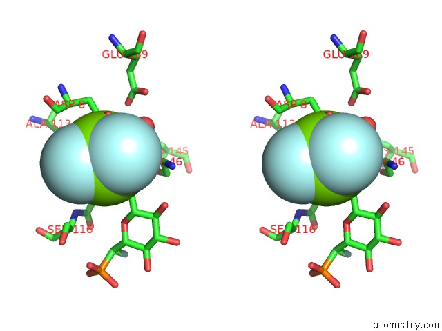
Fluorine binding site 3 out of 4 in 4c4s
Go back to
Fluorine binding site 3 out
of 4 in the Structure of Beta-Phosphoglucomutase in Complex with An Alpha-Fluorophosphonate Analogue of Beta-Glucose-1-Phosphate and Magnesium Trifluoride
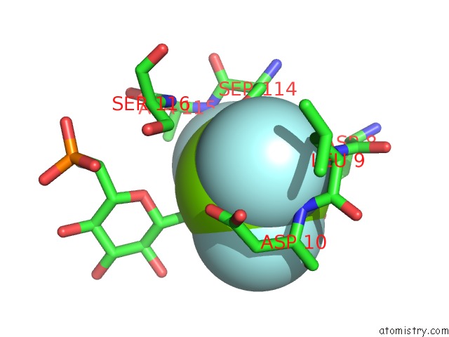
Mono view
Stereo pair view
Mono view
Stereo pair view
A full contact list of Fluorine with other atoms in the F binding
site number 3 of Structure of Beta-Phosphoglucomutase in Complex with An Alpha-Fluorophosphonate Analogue of Beta-Glucose-1-Phosphate and Magnesium Trifluoride within 5.0Å range:
|
Fluorine binding site 4 out of 4 in 4c4s
Go back to
Fluorine binding site 4 out
of 4 in the Structure of Beta-Phosphoglucomutase in Complex with An Alpha-Fluorophosphonate Analogue of Beta-Glucose-1-Phosphate and Magnesium Trifluoride
Mono view
Stereo pair view
Mono view
Stereo pair view
A full contact list of Fluorine with other atoms in the F binding
site number 4 of Structure of Beta-Phosphoglucomutase in Complex with An Alpha-Fluorophosphonate Analogue of Beta-Glucose-1-Phosphate and Magnesium Trifluoride within 5.0Å range:
|
Reference:
Y.Jin,
D.Bhattasali,
E.Pellegrini,
S.M.Forget,
N.J.Baxter,
M.J.Cliff,
M.W.Bowler,
D.L.Jakeman,
G.M.Blackburn,
J.P.Waltho.
Alpha-Fluorophosphonates Reveal How A Phosphomutase Conserves Transition State Conformation Over Hexose Recognition in Its Two-Step Reaction. Proc.Natl.Acad.Sci.Usa V. 111 12384 2014.
ISSN: ISSN 0027-8424
PubMed: 25104750
DOI: 10.1073/PNAS.1402850111
Page generated: Thu Aug 1 00:30:42 2024
ISSN: ISSN 0027-8424
PubMed: 25104750
DOI: 10.1073/PNAS.1402850111
Last articles
Zn in 9MJ5Zn in 9HNW
Zn in 9G0L
Zn in 9FNE
Zn in 9DZN
Zn in 9E0I
Zn in 9D32
Zn in 9DAK
Zn in 8ZXC
Zn in 8ZUF