Fluorine »
PDB 4c6l-4cqj »
4c8b »
Fluorine in PDB 4c8b: Structure of the Kinase Domain of Human RIPK2 in Complex with Ponatinib
Enzymatic activity of Structure of the Kinase Domain of Human RIPK2 in Complex with Ponatinib
All present enzymatic activity of Structure of the Kinase Domain of Human RIPK2 in Complex with Ponatinib:
2.7.10.2; 2.7.11.1;
2.7.10.2; 2.7.11.1;
Protein crystallography data
The structure of Structure of the Kinase Domain of Human RIPK2 in Complex with Ponatinib, PDB code: 4c8b
was solved by
P.Canning,
T.Krojer,
A.Bradley,
P.Mahajan,
S.Goubin,
F.Von Delft,
C.H.Arrowsmith,
A.M.Edwards,
C.Bountra,
A.Bullock,
with X-Ray Crystallography technique. A brief refinement statistics is given in the table below:
| Resolution Low / High (Å) | 54.06 / 2.75 |
| Space group | P 21 21 21 |
| Cell size a, b, c (Å), α, β, γ (°) | 58.735, 86.682, 137.335, 90.00, 90.00, 90.00 |
| R / Rfree (%) | 20.061 / 24.361 |
Fluorine Binding Sites:
The binding sites of Fluorine atom in the Structure of the Kinase Domain of Human RIPK2 in Complex with Ponatinib
(pdb code 4c8b). This binding sites where shown within
5.0 Angstroms radius around Fluorine atom.
In total 6 binding sites of Fluorine where determined in the Structure of the Kinase Domain of Human RIPK2 in Complex with Ponatinib, PDB code: 4c8b:
Jump to Fluorine binding site number: 1; 2; 3; 4; 5; 6;
In total 6 binding sites of Fluorine where determined in the Structure of the Kinase Domain of Human RIPK2 in Complex with Ponatinib, PDB code: 4c8b:
Jump to Fluorine binding site number: 1; 2; 3; 4; 5; 6;
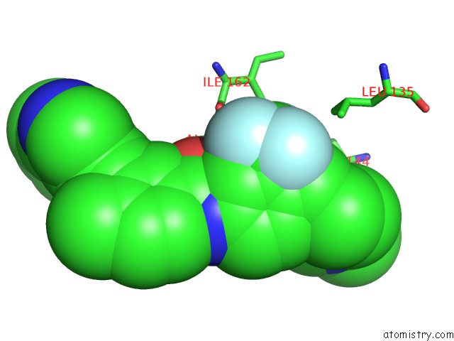
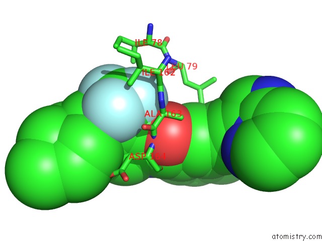
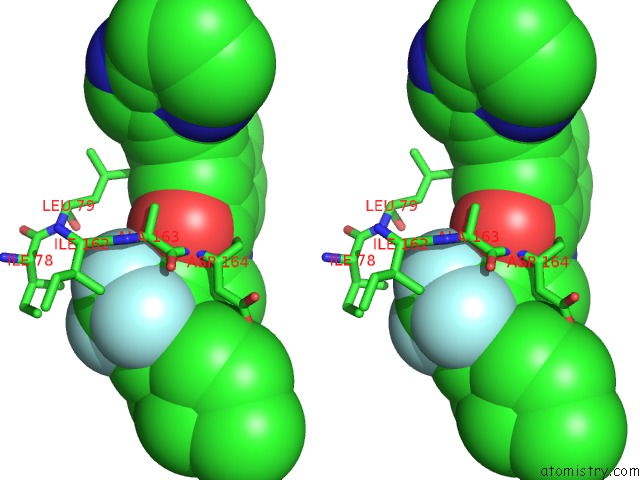
Fluorine binding site 1 out of 6 in 4c8b
Go back to
Fluorine binding site 1 out
of 6 in the Structure of the Kinase Domain of Human RIPK2 in Complex with Ponatinib
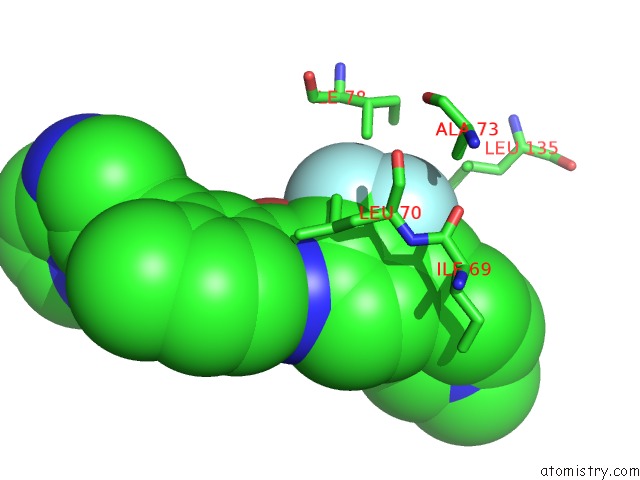
Mono view
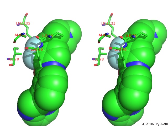
Stereo pair view
Mono view
Stereo pair view
A full contact list of Fluorine with other atoms in the F binding
site number 1 of Structure of the Kinase Domain of Human RIPK2 in Complex with Ponatinib within 5.0Å range:
|
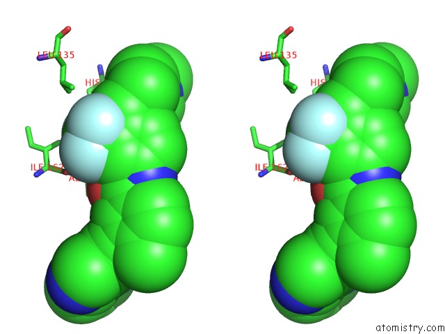
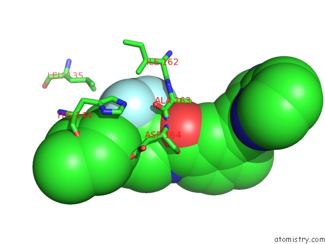
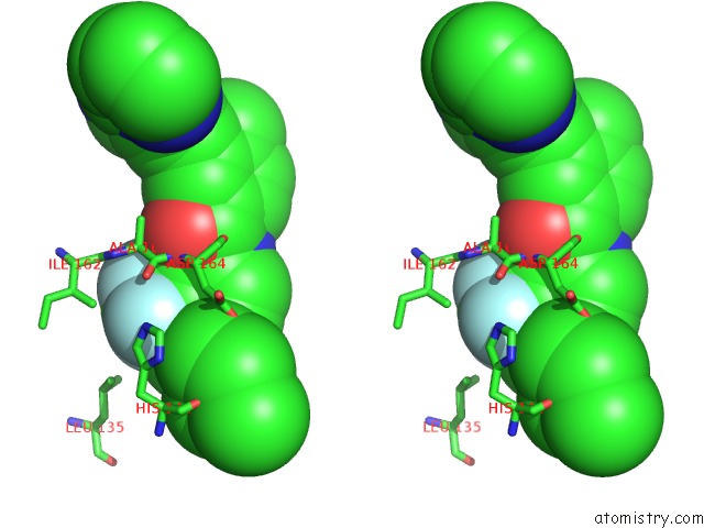
Fluorine binding site 2 out of 6 in 4c8b
Go back to
Fluorine binding site 2 out
of 6 in the Structure of the Kinase Domain of Human RIPK2 in Complex with Ponatinib
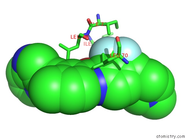
Mono view
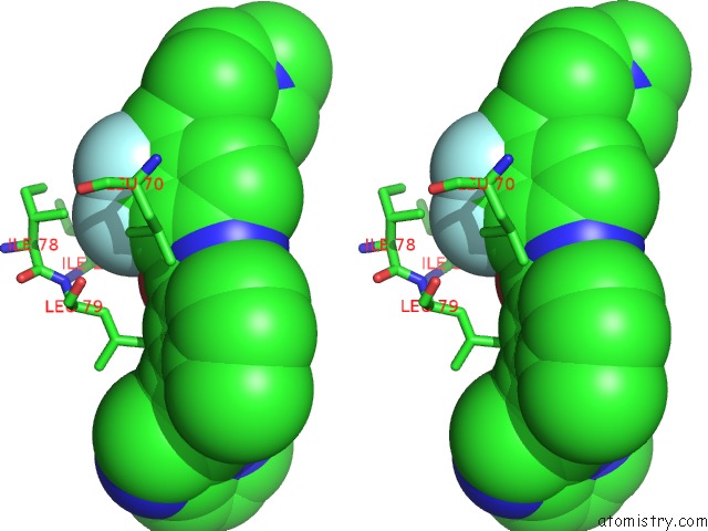
Stereo pair view
Mono view
Stereo pair view
A full contact list of Fluorine with other atoms in the F binding
site number 2 of Structure of the Kinase Domain of Human RIPK2 in Complex with Ponatinib within 5.0Å range:
|
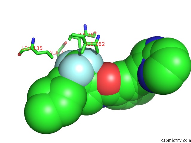
Fluorine binding site 3 out of 6 in 4c8b
Go back to
Fluorine binding site 3 out
of 6 in the Structure of the Kinase Domain of Human RIPK2 in Complex with Ponatinib
Mono view
Stereo pair view
Mono view
Stereo pair view
A full contact list of Fluorine with other atoms in the F binding
site number 3 of Structure of the Kinase Domain of Human RIPK2 in Complex with Ponatinib within 5.0Å range:
|
Fluorine binding site 4 out of 6 in 4c8b
Go back to
Fluorine binding site 4 out
of 6 in the Structure of the Kinase Domain of Human RIPK2 in Complex with Ponatinib
Mono view
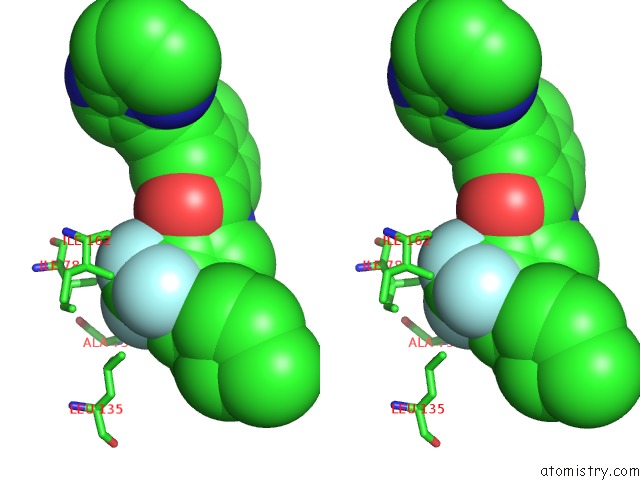
Stereo pair view
Mono view
Stereo pair view
A full contact list of Fluorine with other atoms in the F binding
site number 4 of Structure of the Kinase Domain of Human RIPK2 in Complex with Ponatinib within 5.0Å range:
|
Fluorine binding site 5 out of 6 in 4c8b
Go back to
Fluorine binding site 5 out
of 6 in the Structure of the Kinase Domain of Human RIPK2 in Complex with Ponatinib
Mono view
Stereo pair view
Mono view
Stereo pair view
A full contact list of Fluorine with other atoms in the F binding
site number 5 of Structure of the Kinase Domain of Human RIPK2 in Complex with Ponatinib within 5.0Å range:
|
Fluorine binding site 6 out of 6 in 4c8b
Go back to
Fluorine binding site 6 out
of 6 in the Structure of the Kinase Domain of Human RIPK2 in Complex with Ponatinib
Mono view
Stereo pair view
Mono view
Stereo pair view
A full contact list of Fluorine with other atoms in the F binding
site number 6 of Structure of the Kinase Domain of Human RIPK2 in Complex with Ponatinib within 5.0Å range:
|
Reference:
P.Canning,
A.Bullock.
Structure of the Kinase Domain of Human RIPK2 in Complex with Ponatinib To Be Published.
Page generated: Thu Aug 1 00:40:40 2024
Last articles
F in 4BKZF in 4BFW
F in 4BFX
F in 4BJK
F in 4BJC
F in 4BFU
F in 4BGM
F in 4BFV
F in 4BEW
F in 4BFS