Fluorine »
PDB 4c6l-4cqj »
4c9x »
Fluorine in PDB 4c9x: Crystal Structure of NUDT1 (MTH1) with S-Crizotinib
Enzymatic activity of Crystal Structure of NUDT1 (MTH1) with S-Crizotinib
All present enzymatic activity of Crystal Structure of NUDT1 (MTH1) with S-Crizotinib:
3.6.1.55; 3.6.1.56;
3.6.1.55; 3.6.1.56;
Protein crystallography data
The structure of Crystal Structure of NUDT1 (MTH1) with S-Crizotinib, PDB code: 4c9x
was solved by
J.M.Elkins,
E.Salah,
M.Vollmar,
K.Huber,
G.Superti-Furga,
K.R.Abdul Azeez,
T.Krojer,
F.Von Delft,
C.Bountra,
A.Edwards,
S.Knapp,
with X-Ray Crystallography technique. A brief refinement statistics is given in the table below:
| Resolution Low / High (Å) | 44.71 / 1.20 |
| Space group | P 2 21 21 |
| Cell size a, b, c (Å), α, β, γ (°) | 36.200, 60.020, 67.000, 90.00, 90.00, 90.00 |
| R / Rfree (%) | 14.629 / 18.159 |
Other elements in 4c9x:
The structure of Crystal Structure of NUDT1 (MTH1) with S-Crizotinib also contains other interesting chemical elements:
| Chlorine | (Cl) | 3 atoms |
Fluorine Binding Sites:
The binding sites of Fluorine atom in the Crystal Structure of NUDT1 (MTH1) with S-Crizotinib
(pdb code 4c9x). This binding sites where shown within
5.0 Angstroms radius around Fluorine atom.
In total only one binding site of Fluorine was determined in the Crystal Structure of NUDT1 (MTH1) with S-Crizotinib, PDB code: 4c9x:
In total only one binding site of Fluorine was determined in the Crystal Structure of NUDT1 (MTH1) with S-Crizotinib, PDB code: 4c9x:
Fluorine binding site 1 out of 1 in 4c9x
Go back to
Fluorine binding site 1 out
of 1 in the Crystal Structure of NUDT1 (MTH1) with S-Crizotinib
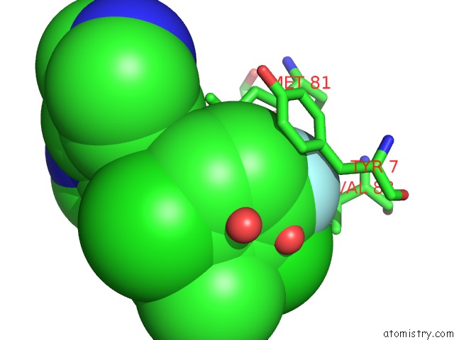
Mono view
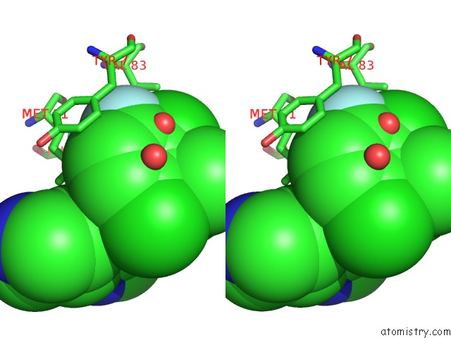
Stereo pair view
Mono view
Stereo pair view
A full contact list of Fluorine with other atoms in the F binding
site number 1 of Crystal Structure of NUDT1 (MTH1) with S-Crizotinib within 5.0Å range:
|
Reference:
K.V.M.Huber,
E.Salah,
B.Radic,
M.Gridling,
J.M.Elkins,
A.Stukalov,
A.Jemth,
C.Gokturk,
K.Sanjiv,
K.Stromberg,
T.Pham,
U.W.Berglund,
J.Colinge,
K.L.Bennett,
J.I.Loizou,
T.Helleday,
S.Knapp,
G.Superti-Furga.
Stereospecific Targeting of MTH1 By (S)-Crizotinib As An Anticancer Strategy. Nature V. 508 222 2014.
ISSN: ISSN 0028-0836
PubMed: 24695225
DOI: 10.1038/NATURE13194
Page generated: Thu Aug 1 00:40:40 2024
ISSN: ISSN 0028-0836
PubMed: 24695225
DOI: 10.1038/NATURE13194
Last articles
Zn in 9J0NZn in 9J0O
Zn in 9J0P
Zn in 9FJX
Zn in 9EKB
Zn in 9C0F
Zn in 9CAH
Zn in 9CH0
Zn in 9CH3
Zn in 9CH1