Fluorine »
PDB 4dbu-4e28 »
4e1v »
Fluorine in PDB 4e1v: X-Ray Structure of the Uridine Phosphorylase From Salmonella Typhimurium in Complex with 5-Fluorouracil at 2.15 A Resolution
Enzymatic activity of X-Ray Structure of the Uridine Phosphorylase From Salmonella Typhimurium in Complex with 5-Fluorouracil at 2.15 A Resolution
All present enzymatic activity of X-Ray Structure of the Uridine Phosphorylase From Salmonella Typhimurium in Complex with 5-Fluorouracil at 2.15 A Resolution:
2.4.2.3;
2.4.2.3;
Protein crystallography data
The structure of X-Ray Structure of the Uridine Phosphorylase From Salmonella Typhimurium in Complex with 5-Fluorouracil at 2.15 A Resolution, PDB code: 4e1v
was solved by
A.A.Lashkov,
S.E.Sotnichenko,
I.I.Prokofev,
A.G.Gabdoulkhakov,
A.M.Mikhailov,
with X-Ray Crystallography technique. A brief refinement statistics is given in the table below:
| Resolution Low / High (Å) | 28.88 / 2.15 |
| Space group | C 1 2 1 |
| Cell size a, b, c (Å), α, β, γ (°) | 158.490, 93.210, 149.970, 90.00, 90.82, 90.00 |
| R / Rfree (%) | 19.4 / 23.8 |
Other elements in 4e1v:
The structure of X-Ray Structure of the Uridine Phosphorylase From Salmonella Typhimurium in Complex with 5-Fluorouracil at 2.15 A Resolution also contains other interesting chemical elements:
| Potassium | (K) | 3 atoms |
Fluorine Binding Sites:
The binding sites of Fluorine atom in the X-Ray Structure of the Uridine Phosphorylase From Salmonella Typhimurium in Complex with 5-Fluorouracil at 2.15 A Resolution
(pdb code 4e1v). This binding sites where shown within
5.0 Angstroms radius around Fluorine atom.
In total 8 binding sites of Fluorine where determined in the X-Ray Structure of the Uridine Phosphorylase From Salmonella Typhimurium in Complex with 5-Fluorouracil at 2.15 A Resolution, PDB code: 4e1v:
Jump to Fluorine binding site number: 1; 2; 3; 4; 5; 6; 7; 8;
In total 8 binding sites of Fluorine where determined in the X-Ray Structure of the Uridine Phosphorylase From Salmonella Typhimurium in Complex with 5-Fluorouracil at 2.15 A Resolution, PDB code: 4e1v:
Jump to Fluorine binding site number: 1; 2; 3; 4; 5; 6; 7; 8;
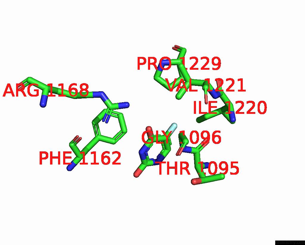
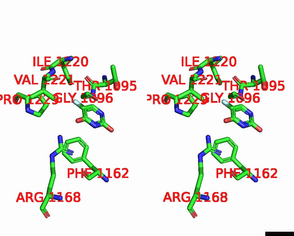
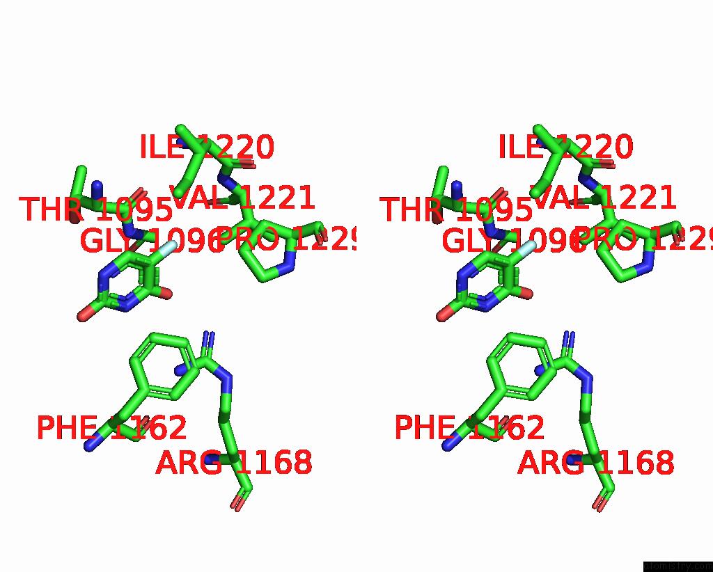
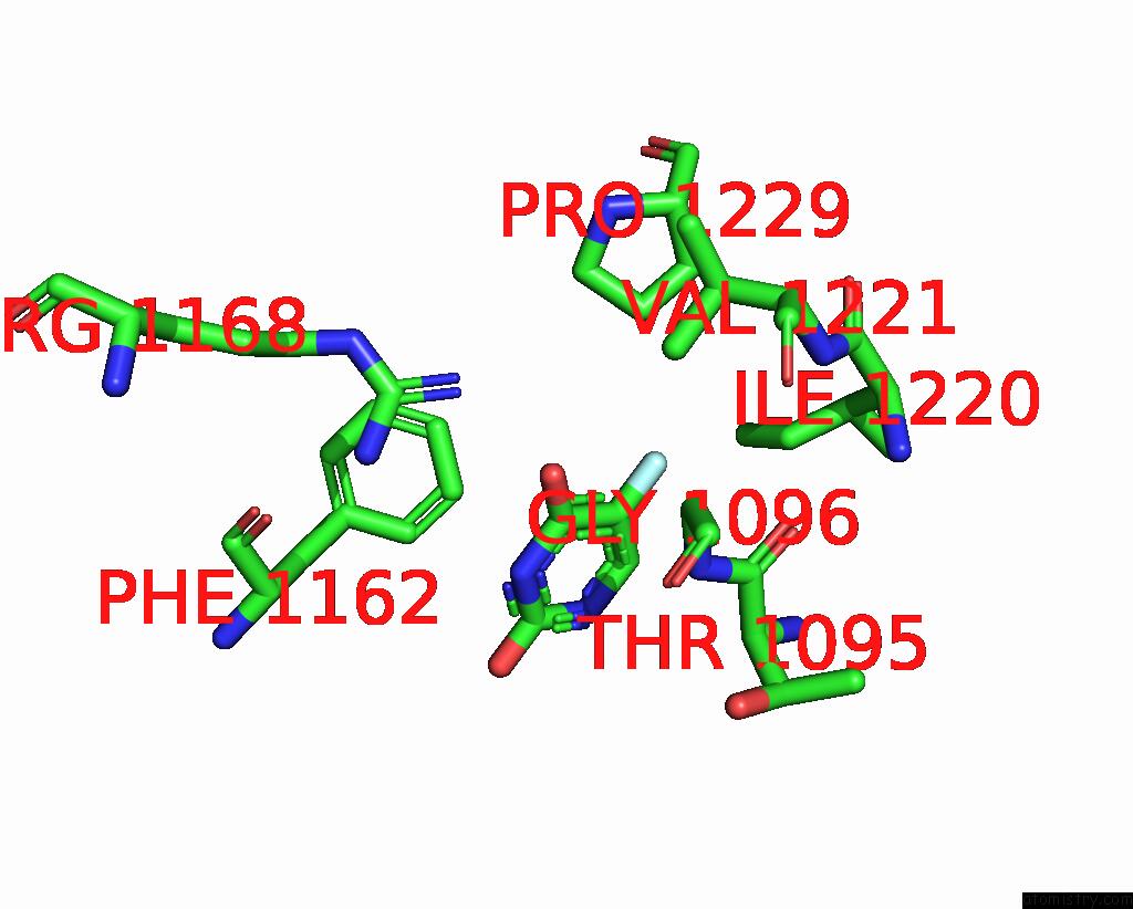
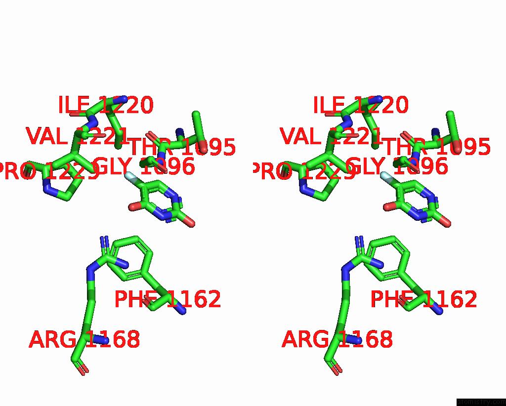
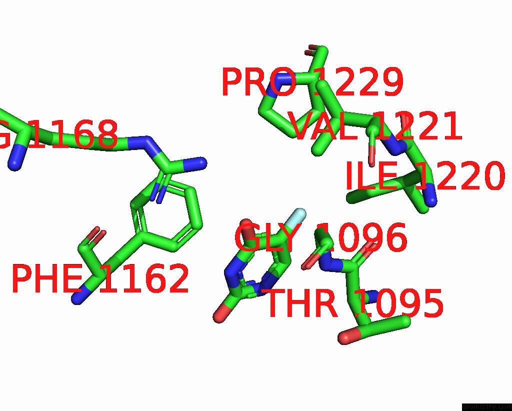
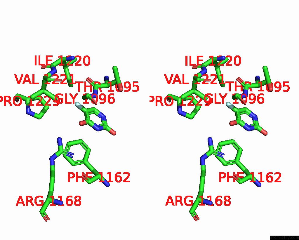
Fluorine binding site 1 out of 8 in 4e1v
Go back to
Fluorine binding site 1 out
of 8 in the X-Ray Structure of the Uridine Phosphorylase From Salmonella Typhimurium in Complex with 5-Fluorouracil at 2.15 A Resolution
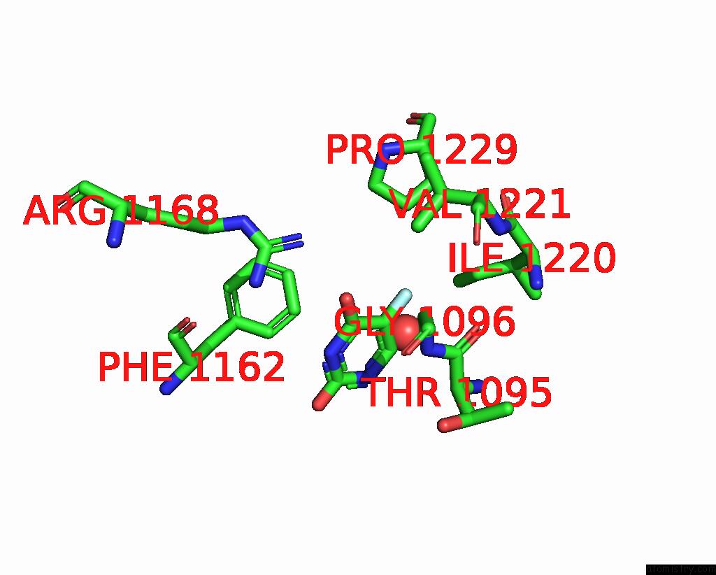
Mono view
Stereo pair view
Mono view
Stereo pair view
A full contact list of Fluorine with other atoms in the F binding
site number 1 of X-Ray Structure of the Uridine Phosphorylase From Salmonella Typhimurium in Complex with 5-Fluorouracil at 2.15 A Resolution within 5.0Å range:
|
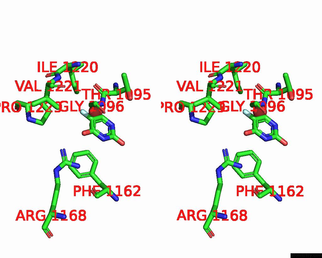
Fluorine binding site 2 out of 8 in 4e1v
Go back to
Fluorine binding site 2 out
of 8 in the X-Ray Structure of the Uridine Phosphorylase From Salmonella Typhimurium in Complex with 5-Fluorouracil at 2.15 A Resolution

Mono view
Stereo pair view
Mono view
Stereo pair view
A full contact list of Fluorine with other atoms in the F binding
site number 2 of X-Ray Structure of the Uridine Phosphorylase From Salmonella Typhimurium in Complex with 5-Fluorouracil at 2.15 A Resolution within 5.0Å range:
|
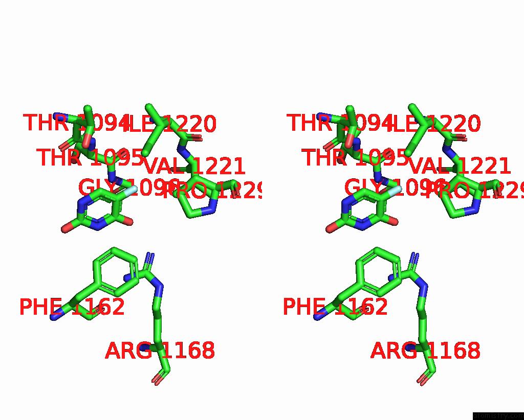
Fluorine binding site 3 out of 8 in 4e1v
Go back to
Fluorine binding site 3 out
of 8 in the X-Ray Structure of the Uridine Phosphorylase From Salmonella Typhimurium in Complex with 5-Fluorouracil at 2.15 A Resolution
Mono view

Stereo pair view
Mono view
Stereo pair view
A full contact list of Fluorine with other atoms in the F binding
site number 3 of X-Ray Structure of the Uridine Phosphorylase From Salmonella Typhimurium in Complex with 5-Fluorouracil at 2.15 A Resolution within 5.0Å range:
|
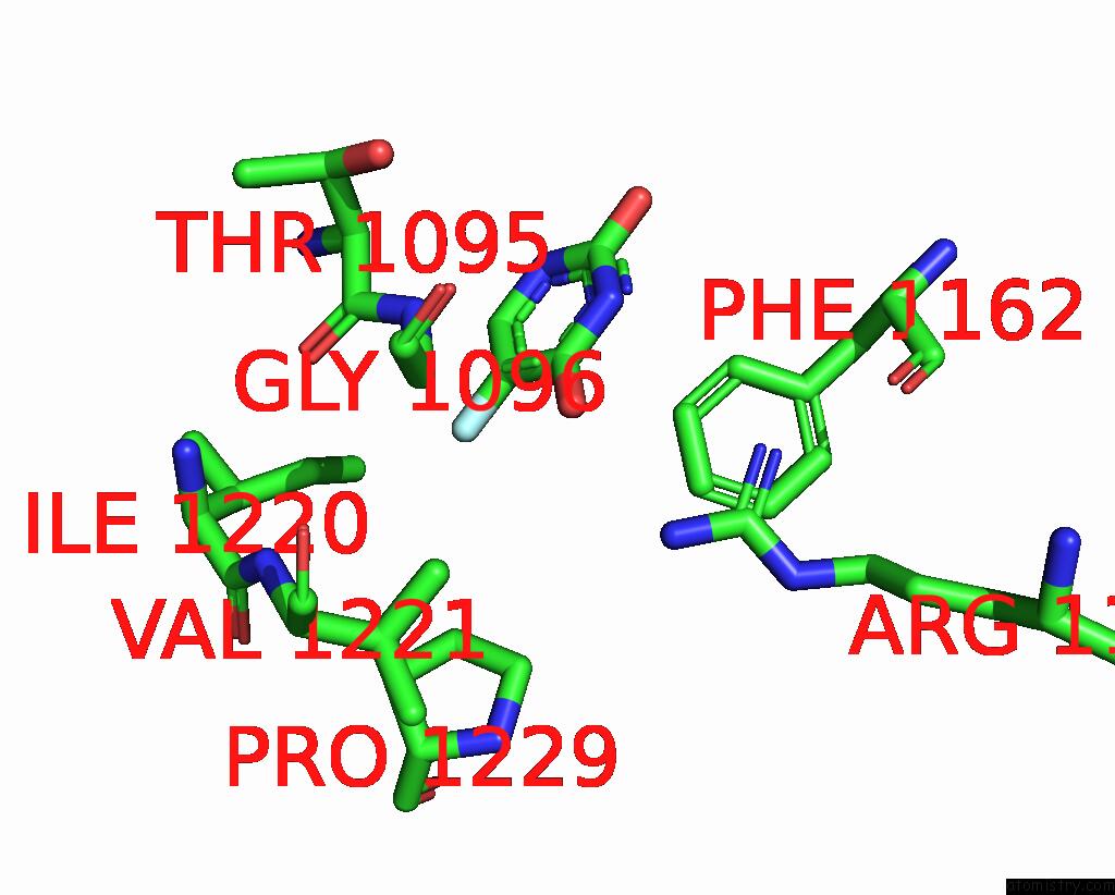
Fluorine binding site 4 out of 8 in 4e1v
Go back to
Fluorine binding site 4 out
of 8 in the X-Ray Structure of the Uridine Phosphorylase From Salmonella Typhimurium in Complex with 5-Fluorouracil at 2.15 A Resolution
Mono view
Stereo pair view
Mono view
Stereo pair view
A full contact list of Fluorine with other atoms in the F binding
site number 4 of X-Ray Structure of the Uridine Phosphorylase From Salmonella Typhimurium in Complex with 5-Fluorouracil at 2.15 A Resolution within 5.0Å range:
|
Fluorine binding site 5 out of 8 in 4e1v
Go back to
Fluorine binding site 5 out
of 8 in the X-Ray Structure of the Uridine Phosphorylase From Salmonella Typhimurium in Complex with 5-Fluorouracil at 2.15 A Resolution

Mono view
Stereo pair view
Mono view
Stereo pair view
A full contact list of Fluorine with other atoms in the F binding
site number 5 of X-Ray Structure of the Uridine Phosphorylase From Salmonella Typhimurium in Complex with 5-Fluorouracil at 2.15 A Resolution within 5.0Å range:
|
Fluorine binding site 6 out of 8 in 4e1v
Go back to
Fluorine binding site 6 out
of 8 in the X-Ray Structure of the Uridine Phosphorylase From Salmonella Typhimurium in Complex with 5-Fluorouracil at 2.15 A Resolution
Mono view
Stereo pair view
Mono view
Stereo pair view
A full contact list of Fluorine with other atoms in the F binding
site number 6 of X-Ray Structure of the Uridine Phosphorylase From Salmonella Typhimurium in Complex with 5-Fluorouracil at 2.15 A Resolution within 5.0Å range:
|
Fluorine binding site 7 out of 8 in 4e1v
Go back to
Fluorine binding site 7 out
of 8 in the X-Ray Structure of the Uridine Phosphorylase From Salmonella Typhimurium in Complex with 5-Fluorouracil at 2.15 A Resolution
Mono view
Stereo pair view
Mono view
Stereo pair view
A full contact list of Fluorine with other atoms in the F binding
site number 7 of X-Ray Structure of the Uridine Phosphorylase From Salmonella Typhimurium in Complex with 5-Fluorouracil at 2.15 A Resolution within 5.0Å range:
|
Fluorine binding site 8 out of 8 in 4e1v
Go back to
Fluorine binding site 8 out
of 8 in the X-Ray Structure of the Uridine Phosphorylase From Salmonella Typhimurium in Complex with 5-Fluorouracil at 2.15 A Resolution

Mono view

Stereo pair view
Mono view
Stereo pair view
A full contact list of Fluorine with other atoms in the F binding
site number 8 of X-Ray Structure of the Uridine Phosphorylase From Salmonella Typhimurium in Complex with 5-Fluorouracil at 2.15 A Resolution within 5.0Å range:
|
Reference:
A.A.Lashkov,
S.E.Sotnichenko,
I.I.Prokofiev,
A.G.Gabdulkhakov,
I.I.Agapov,
A.A.Shtil,
C.Betzel,
A.S.Mironov,
A.M.Mikhailov.
X-Ray Structure of Salmonella Typhimurium Uridine Phosphorylase Complexed with 5-Fluorouracil and Molecular Modelling of the Complex of 5-Fluorouracil with Uridine Phosphorylase From Vibrio Cholerae. Acta Crystallogr.,Sect.D V. 68 968 2012.
ISSN: ISSN 0907-4449
PubMed: 22868762
DOI: 10.1107/S090744491201815X
Page generated: Thu Aug 1 01:10:15 2024
ISSN: ISSN 0907-4449
PubMed: 22868762
DOI: 10.1107/S090744491201815X
Last articles
Zn in 9MJ5Zn in 9HNW
Zn in 9G0L
Zn in 9FNE
Zn in 9DZN
Zn in 9E0I
Zn in 9D32
Zn in 9DAK
Zn in 8ZXC
Zn in 8ZUF