Fluorine »
PDB 4e3n-4f9m »
4ear »
Fluorine in PDB 4ear: Crystal Structure of Purine Nucleoside Phosphorylase (W16Y, W94Y, W178Y, H257W) Mutant From Human Complexed with Dadme-Immg and Phosphate
Enzymatic activity of Crystal Structure of Purine Nucleoside Phosphorylase (W16Y, W94Y, W178Y, H257W) Mutant From Human Complexed with Dadme-Immg and Phosphate
All present enzymatic activity of Crystal Structure of Purine Nucleoside Phosphorylase (W16Y, W94Y, W178Y, H257W) Mutant From Human Complexed with Dadme-Immg and Phosphate:
2.4.2.1;
2.4.2.1;
Protein crystallography data
The structure of Crystal Structure of Purine Nucleoside Phosphorylase (W16Y, W94Y, W178Y, H257W) Mutant From Human Complexed with Dadme-Immg and Phosphate, PDB code: 4ear
was solved by
A.M.Haapalainen,
M.C.Ho,
J.J.Suarez,
S.C.Almo,
V.L.Schramm,
with X-Ray Crystallography technique. A brief refinement statistics is given in the table below:
| Resolution Low / High (Å) | 41.24 / 1.70 |
| Space group | P 21 21 21 |
| Cell size a, b, c (Å), α, β, γ (°) | 55.916, 131.100, 137.680, 90.00, 90.00, 90.00 |
| R / Rfree (%) | 17.5 / 20.3 |
Fluorine Binding Sites:
The binding sites of Fluorine atom in the Crystal Structure of Purine Nucleoside Phosphorylase (W16Y, W94Y, W178Y, H257W) Mutant From Human Complexed with Dadme-Immg and Phosphate
(pdb code 4ear). This binding sites where shown within
5.0 Angstroms radius around Fluorine atom.
In total 3 binding sites of Fluorine where determined in the Crystal Structure of Purine Nucleoside Phosphorylase (W16Y, W94Y, W178Y, H257W) Mutant From Human Complexed with Dadme-Immg and Phosphate, PDB code: 4ear:
Jump to Fluorine binding site number: 1; 2; 3;
In total 3 binding sites of Fluorine where determined in the Crystal Structure of Purine Nucleoside Phosphorylase (W16Y, W94Y, W178Y, H257W) Mutant From Human Complexed with Dadme-Immg and Phosphate, PDB code: 4ear:
Jump to Fluorine binding site number: 1; 2; 3;
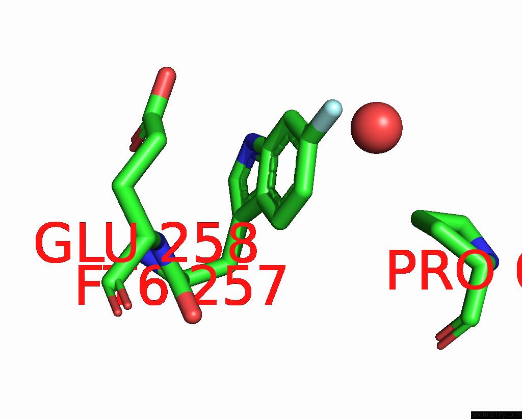
Fluorine binding site 1 out of 3 in 4ear
Go back to
Fluorine binding site 1 out
of 3 in the Crystal Structure of Purine Nucleoside Phosphorylase (W16Y, W94Y, W178Y, H257W) Mutant From Human Complexed with Dadme-Immg and Phosphate

Mono view

Stereo pair view
Mono view
Stereo pair view
A full contact list of Fluorine with other atoms in the F binding
site number 1 of Crystal Structure of Purine Nucleoside Phosphorylase (W16Y, W94Y, W178Y, H257W) Mutant From Human Complexed with Dadme-Immg and Phosphate within 5.0Å range:
|
Fluorine binding site 2 out of 3 in 4ear
Go back to
Fluorine binding site 2 out
of 3 in the Crystal Structure of Purine Nucleoside Phosphorylase (W16Y, W94Y, W178Y, H257W) Mutant From Human Complexed with Dadme-Immg and Phosphate
Mono view

Stereo pair view
Mono view
Stereo pair view
A full contact list of Fluorine with other atoms in the F binding
site number 2 of Crystal Structure of Purine Nucleoside Phosphorylase (W16Y, W94Y, W178Y, H257W) Mutant From Human Complexed with Dadme-Immg and Phosphate within 5.0Å range:
|
Fluorine binding site 3 out of 3 in 4ear
Go back to
Fluorine binding site 3 out
of 3 in the Crystal Structure of Purine Nucleoside Phosphorylase (W16Y, W94Y, W178Y, H257W) Mutant From Human Complexed with Dadme-Immg and Phosphate

Mono view

Stereo pair view
Mono view
Stereo pair view
A full contact list of Fluorine with other atoms in the F binding
site number 3 of Crystal Structure of Purine Nucleoside Phosphorylase (W16Y, W94Y, W178Y, H257W) Mutant From Human Complexed with Dadme-Immg and Phosphate within 5.0Å range:
|
Reference:
J.Suarez,
A.M.Haapalainen,
S.M.Cahill,
M.C.Ho,
F.Yan,
S.C.Almo,
V.L.Schramm.
Catalytic Site Conformations in Human Pnp By (19)F-uc(Nmr) and Crystallography. Chem.Biol. V. 20 212 2013.
ISSN: ISSN 1074-5521
PubMed: 23438750
DOI: 10.1016/J.CHEMBIOL.2013.01.009
Page generated: Mon Jul 14 21:23:56 2025
ISSN: ISSN 1074-5521
PubMed: 23438750
DOI: 10.1016/J.CHEMBIOL.2013.01.009
Last articles
F in 4WNKF in 4WI1
F in 4WMZ
F in 4WMV
F in 4WM7
F in 4WLB
F in 4WKQ
F in 4WHQ
F in 4WGI
F in 4WF5