Fluorine »
PDB 4fxq-4goa »
4glx »
Fluorine in PDB 4glx: Dna Ligase A in Complex with Inhibitor
Enzymatic activity of Dna Ligase A in Complex with Inhibitor
All present enzymatic activity of Dna Ligase A in Complex with Inhibitor:
6.5.1.2;
6.5.1.2;
Protein crystallography data
The structure of Dna Ligase A in Complex with Inhibitor, PDB code: 4glx
was solved by
L.Prade,
R.Lange,
N.Tidten-Luksch,
A.Chambovey,
with X-Ray Crystallography technique. A brief refinement statistics is given in the table below:
| Resolution Low / High (Å) | 19.73 / 1.90 |
| Space group | C 1 2 1 |
| Cell size a, b, c (Å), α, β, γ (°) | 110.990, 100.140, 86.570, 90.00, 105.50, 90.00 |
| R / Rfree (%) | 20.1 / 25 |
Other elements in 4glx:
The structure of Dna Ligase A in Complex with Inhibitor also contains other interesting chemical elements:
| Bromine | (Br) | 1 atom |
| Zinc | (Zn) | 1 atom |
Fluorine Binding Sites:
The binding sites of Fluorine atom in the Dna Ligase A in Complex with Inhibitor
(pdb code 4glx). This binding sites where shown within
5.0 Angstroms radius around Fluorine atom.
In total 3 binding sites of Fluorine where determined in the Dna Ligase A in Complex with Inhibitor, PDB code: 4glx:
Jump to Fluorine binding site number: 1; 2; 3;
In total 3 binding sites of Fluorine where determined in the Dna Ligase A in Complex with Inhibitor, PDB code: 4glx:
Jump to Fluorine binding site number: 1; 2; 3;
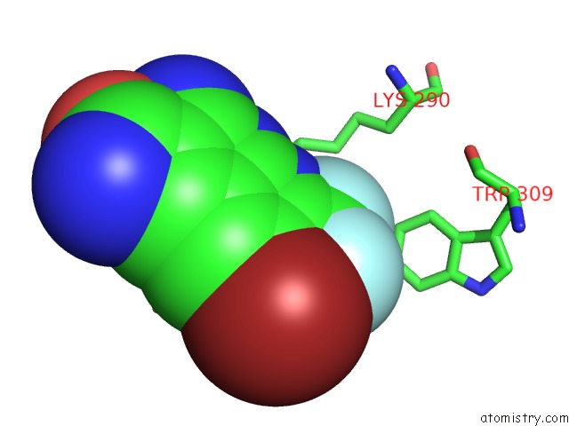
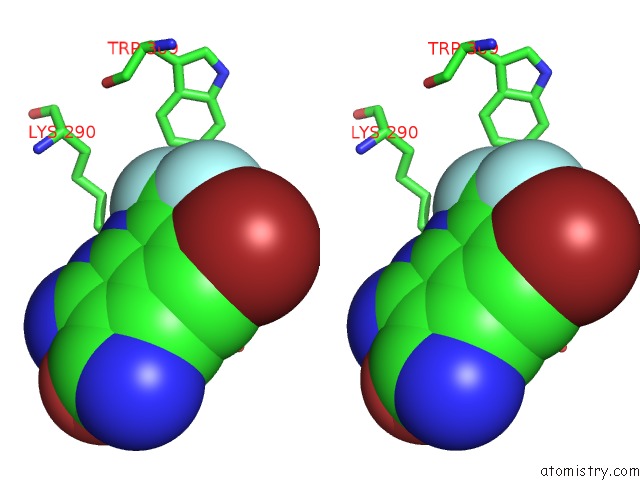
Fluorine binding site 1 out of 3 in 4glx
Go back to
Fluorine binding site 1 out
of 3 in the Dna Ligase A in Complex with Inhibitor

Mono view

Stereo pair view
Mono view
Stereo pair view
A full contact list of Fluorine with other atoms in the F binding
site number 1 of Dna Ligase A in Complex with Inhibitor within 5.0Å range:
|
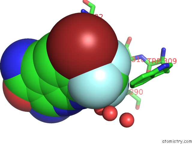
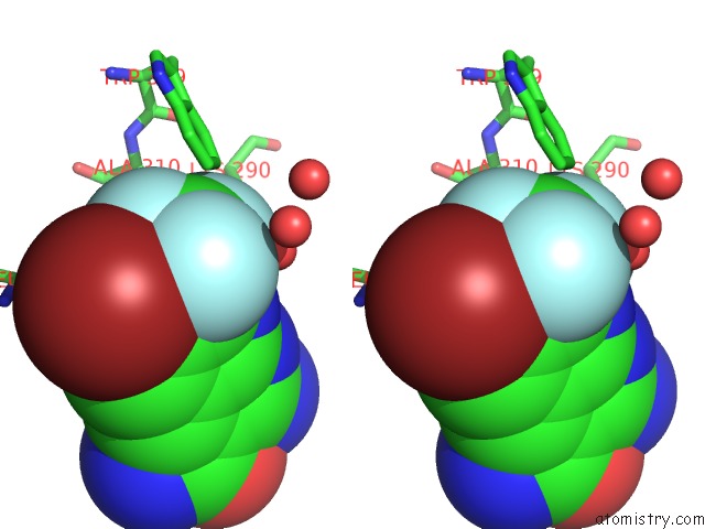
Fluorine binding site 2 out of 3 in 4glx
Go back to
Fluorine binding site 2 out
of 3 in the Dna Ligase A in Complex with Inhibitor
Mono view
Stereo pair view
Mono view
Stereo pair view
A full contact list of Fluorine with other atoms in the F binding
site number 2 of Dna Ligase A in Complex with Inhibitor within 5.0Å range:
|
Fluorine binding site 3 out of 3 in 4glx
Go back to
Fluorine binding site 3 out
of 3 in the Dna Ligase A in Complex with Inhibitor
Mono view
Stereo pair view
Mono view
Stereo pair view
A full contact list of Fluorine with other atoms in the F binding
site number 3 of Dna Ligase A in Complex with Inhibitor within 5.0Å range:
|
Reference:
J.P.Surivet,
R.Lange,
C.Hubschwerlen,
W.Keck,
J.L.Specklin,
D.Ritz,
D.Bur,
H.Locher,
P.Seiler,
D.S.Strasser,
L.Prade,
C.Kohl,
C.Schmitt,
G.Chapoux,
E.Ilhan,
N.Ekambaram,
A.Athanasiou,
A.Knezevic,
D.Sabato,
A.Chambovey,
M.Gaertner,
M.Enderlin,
M.Boehme,
V.Sippel,
P.Wyss.
Structure-Guided Design, Synthesis and Biological Evaluation of Novel Dna Ligase Inhibitors with in Vitro and in Vivo Anti-Staphylococcal Activity. Bioorg.Med.Chem.Lett. V. 22 6705 2012.
ISSN: ISSN 0960-894X
PubMed: 23006603
DOI: 10.1016/J.BMCL.2012.08.094
Page generated: Thu Aug 1 01:53:11 2024
ISSN: ISSN 0960-894X
PubMed: 23006603
DOI: 10.1016/J.BMCL.2012.08.094
Last articles
Zn in 9MJ5Zn in 9HNW
Zn in 9G0L
Zn in 9FNE
Zn in 9DZN
Zn in 9E0I
Zn in 9D32
Zn in 9DAK
Zn in 8ZXC
Zn in 8ZUF