Fluorine »
PDB 4hxn-4ijh »
4i23 »
Fluorine in PDB 4i23: Crystal Structure of the Wild-Type Egfr Kinase Domain in Complex with Dacomitinib (Soaked)
Enzymatic activity of Crystal Structure of the Wild-Type Egfr Kinase Domain in Complex with Dacomitinib (Soaked)
All present enzymatic activity of Crystal Structure of the Wild-Type Egfr Kinase Domain in Complex with Dacomitinib (Soaked):
2.7.10.1;
2.7.10.1;
Protein crystallography data
The structure of Crystal Structure of the Wild-Type Egfr Kinase Domain in Complex with Dacomitinib (Soaked), PDB code: 4i23
was solved by
K.S.Gajiwala,
J.Feng,
R.Ferre,
K.Ryan,
O.Brodsky,
A.Stewart,
with X-Ray Crystallography technique. A brief refinement statistics is given in the table below:
| Resolution Low / High (Å) | 19.92 / 2.80 |
| Space group | I 2 3 |
| Cell size a, b, c (Å), α, β, γ (°) | 146.342, 146.342, 146.342, 90.00, 90.00, 90.00 |
| R / Rfree (%) | 47.6 / 29.8 |
Other elements in 4i23:
The structure of Crystal Structure of the Wild-Type Egfr Kinase Domain in Complex with Dacomitinib (Soaked) also contains other interesting chemical elements:
| Chlorine | (Cl) | 1 atom |
Fluorine Binding Sites:
The binding sites of Fluorine atom in the Crystal Structure of the Wild-Type Egfr Kinase Domain in Complex with Dacomitinib (Soaked)
(pdb code 4i23). This binding sites where shown within
5.0 Angstroms radius around Fluorine atom.
In total only one binding site of Fluorine was determined in the Crystal Structure of the Wild-Type Egfr Kinase Domain in Complex with Dacomitinib (Soaked), PDB code: 4i23:
In total only one binding site of Fluorine was determined in the Crystal Structure of the Wild-Type Egfr Kinase Domain in Complex with Dacomitinib (Soaked), PDB code: 4i23:
Fluorine binding site 1 out of 1 in 4i23
Go back to
Fluorine binding site 1 out
of 1 in the Crystal Structure of the Wild-Type Egfr Kinase Domain in Complex with Dacomitinib (Soaked)
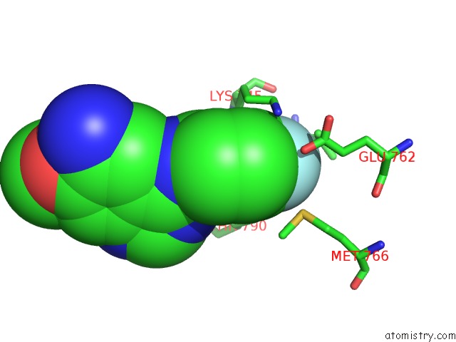
Mono view
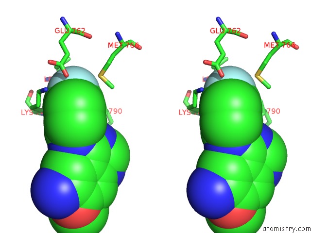
Stereo pair view
Mono view
Stereo pair view
A full contact list of Fluorine with other atoms in the F binding
site number 1 of Crystal Structure of the Wild-Type Egfr Kinase Domain in Complex with Dacomitinib (Soaked) within 5.0Å range:
|
Reference:
K.S.Gajiwala,
J.Feng,
R.Ferre,
K.Ryan,
O.Brodsky,
S.Weinrich,
J.C.Kath,
A.Stewart.
Insights Into the Aberrant Activity of Mutant Egfr Kinase Domain and Drug Recognition. Structure V. 21 209 2013.
ISSN: ISSN 0969-2126
PubMed: 23273428
DOI: 10.1016/J.STR.2012.11.014
Page generated: Thu Aug 1 02:14:13 2024
ISSN: ISSN 0969-2126
PubMed: 23273428
DOI: 10.1016/J.STR.2012.11.014
Last articles
Zn in 9J0NZn in 9J0O
Zn in 9J0P
Zn in 9FJX
Zn in 9EKB
Zn in 9C0F
Zn in 9CAH
Zn in 9CH0
Zn in 9CH3
Zn in 9CH1