Fluorine in PDB 4iah: Crystal Structure of Bay 60-2770 Bound C139A H-Nox Domain with S- Nitrosylated Conserved C122
Enzymatic activity of Crystal Structure of Bay 60-2770 Bound C139A H-Nox Domain with S- Nitrosylated Conserved C122
All present enzymatic activity of Crystal Structure of Bay 60-2770 Bound C139A H-Nox Domain with S- Nitrosylated Conserved C122:
4.6.1.2;
Protein crystallography data
The structure of Crystal Structure of Bay 60-2770 Bound C139A H-Nox Domain with S- Nitrosylated Conserved C122, PDB code: 4iah
was solved by
V.Kumar,
F.Van Den Akker,
with X-Ray Crystallography technique. A brief refinement statistics is given in the table below:
Resolution Low / High (Å)
|
86.69 /
2.80
|
Space group
|
P 21 3
|
Cell size a, b, c (Å), α, β, γ (°)
|
122.573,
122.573,
122.573,
90.00,
90.00,
90.00
|
R / Rfree (%)
|
16.3 /
21.8
|
Fluorine Binding Sites:
The binding sites of Fluorine atom in the Crystal Structure of Bay 60-2770 Bound C139A H-Nox Domain with S- Nitrosylated Conserved C122
(pdb code 4iah). This binding sites where shown within
5.0 Angstroms radius around Fluorine atom.
In total 8 binding sites of Fluorine where determined in the
Crystal Structure of Bay 60-2770 Bound C139A H-Nox Domain with S- Nitrosylated Conserved C122, PDB code: 4iah:
Jump to Fluorine binding site number:
1;
2;
3;
4;
5;
6;
7;
8;
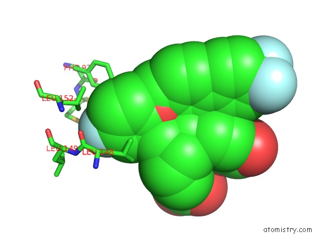
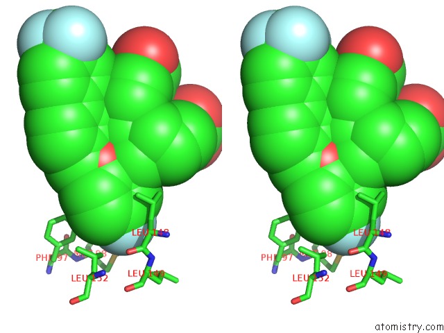
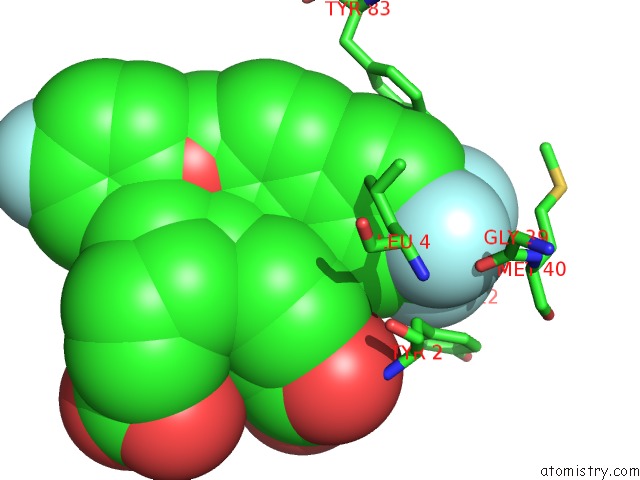
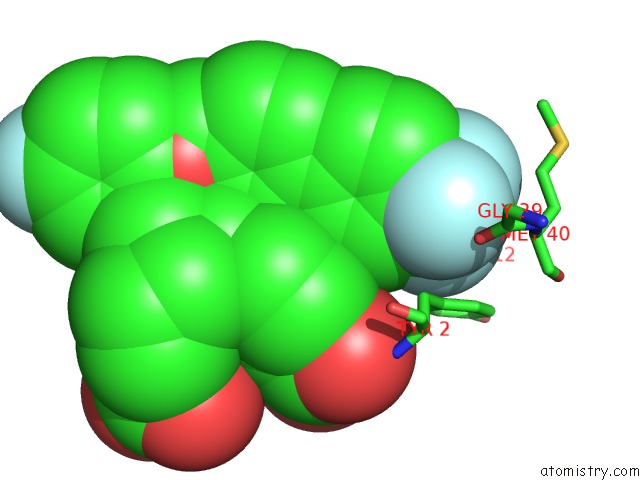
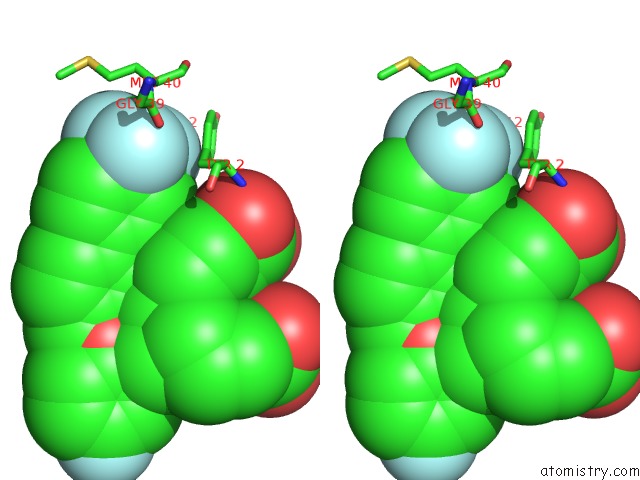
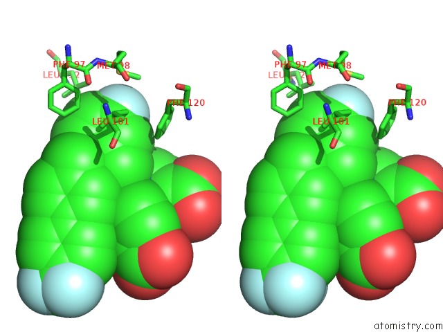
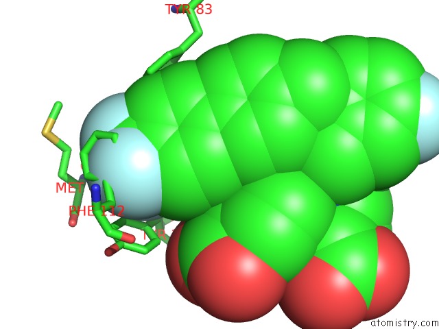
Fluorine binding site 1 out
of 8 in 4iah
Go back to
Fluorine Binding Sites List in 4iah
Fluorine binding site 1 out
of 8 in the Crystal Structure of Bay 60-2770 Bound C139A H-Nox Domain with S- Nitrosylated Conserved C122
 Mono view
 Stereo pair view
|
A full contact list of Fluorine with other atoms in the F binding
site number 1 of Crystal Structure of Bay 60-2770 Bound C139A H-Nox Domain with S- Nitrosylated Conserved C122 within 5.0Å range:
probe
|
atom
|
residue
|
distance (Å)
|
B
|
Occ
|
A:F201
b:27.5
occ:1.00
|
FAA
|
A:1DX201
|
0.0
|
27.5
|
1.0
|
CAC
|
A:1DX201
|
1.3
|
27.8
|
1.0
|
CAB
|
A:1DX201
|
2.4
|
28.1
|
1.0
|
CAD
|
A:1DX201
|
2.4
|
27.6
|
1.0
|
CD1
|
A:LEU152
|
3.5
|
29.1
|
1.0
|
CAE
|
A:1DX201
|
3.6
|
28.1
|
1.0
|
CBN
|
A:1DX201
|
3.6
|
28.0
|
1.0
|
CB
|
A:LEU148
|
3.6
|
26.6
|
1.0
|
CD1
|
A:LEU101
|
3.9
|
34.4
|
1.0
|
SD
|
A:MET98
|
4.0
|
34.2
|
1.0
|
CBO
|
A:1DX201
|
4.1
|
28.4
|
1.0
|
CE2
|
A:PHE97
|
4.2
|
33.2
|
1.0
|
CG
|
A:LEU148
|
4.3
|
26.4
|
1.0
|
CD1
|
A:LEU148
|
4.3
|
26.1
|
1.0
|
CG
|
A:LEU152
|
4.4
|
29.4
|
1.0
|
CD2
|
A:LEU148
|
4.4
|
26.1
|
1.0
|
C
|
A:LEU148
|
4.5
|
26.8
|
1.0
|
CE
|
A:MET98
|
4.6
|
34.4
|
1.0
|
O
|
A:LEU148
|
4.7
|
26.6
|
1.0
|
CD2
|
A:LEU101
|
4.7
|
34.3
|
1.0
|
CA
|
A:LEU148
|
4.7
|
26.6
|
1.0
|
CG
|
A:LEU101
|
4.8
|
34.5
|
1.0
|
N
|
A:LEU149
|
4.8
|
27.1
|
1.0
|
CBA
|
A:1DX201
|
4.9
|
28.1
|
1.0
|
CD2
|
A:PHE97
|
4.9
|
33.1
|
1.0
|
|
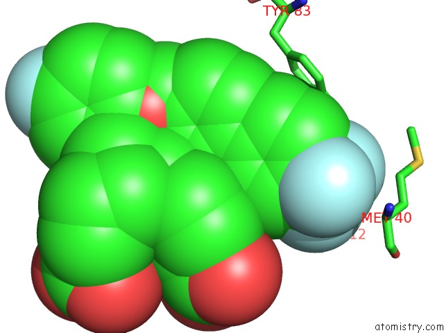
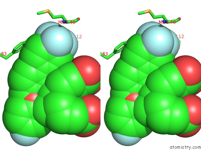
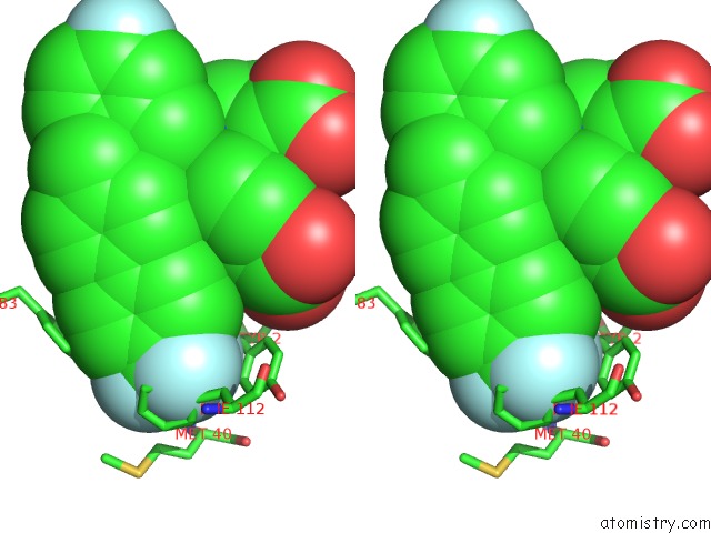
Fluorine binding site 2 out
of 8 in 4iah
Go back to
Fluorine Binding Sites List in 4iah
Fluorine binding site 2 out
of 8 in the Crystal Structure of Bay 60-2770 Bound C139A H-Nox Domain with S- Nitrosylated Conserved C122
 Mono view
 Stereo pair view
|
A full contact list of Fluorine with other atoms in the F binding
site number 2 of Crystal Structure of Bay 60-2770 Bound C139A H-Nox Domain with S- Nitrosylated Conserved C122 within 5.0Å range:
probe
|
atom
|
residue
|
distance (Å)
|
B
|
Occ
|
A:F201
b:33.8
occ:1.00
|
FAE
|
A:1DX201
|
0.0
|
33.8
|
1.0
|
CAI
|
A:1DX201
|
1.3
|
33.6
|
1.0
|
FAJ
|
A:1DX201
|
2.2
|
33.4
|
1.0
|
FAK
|
A:1DX201
|
2.2
|
33.9
|
1.0
|
CAH
|
A:1DX201
|
2.3
|
33.1
|
1.0
|
CZ
|
A:PHE112
|
2.5
|
44.1
|
1.0
|
CE2
|
A:PHE112
|
2.7
|
44.0
|
1.0
|
CAG
|
A:1DX201
|
2.8
|
32.7
|
1.0
|
CE1
|
A:PHE112
|
2.9
|
44.0
|
1.0
|
CD2
|
A:PHE112
|
3.1
|
43.7
|
1.0
|
CD1
|
A:PHE112
|
3.3
|
43.8
|
1.0
|
OH
|
A:TYR83
|
3.4
|
32.9
|
1.0
|
CG
|
A:PHE112
|
3.4
|
43.3
|
1.0
|
CZD
|
A:1DX201
|
3.5
|
32.7
|
1.0
|
CG
|
A:MET40
|
4.1
|
38.8
|
1.0
|
CAJ
|
A:1DX201
|
4.1
|
32.3
|
1.0
|
CZ
|
A:TYR83
|
4.5
|
32.6
|
1.0
|
CB
|
A:PHE112
|
4.6
|
43.0
|
1.0
|
CAK
|
A:1DX201
|
4.6
|
32.4
|
1.0
|
CB
|
A:MET40
|
4.7
|
38.9
|
1.0
|
CA
|
A:MET40
|
4.8
|
38.9
|
1.0
|
SD
|
A:MET40
|
4.8
|
38.5
|
1.0
|
CE1
|
A:TYR83
|
4.9
|
32.5
|
1.0
|
CE
|
A:MET40
|
4.9
|
38.5
|
1.0
|
CBI
|
A:1DX201
|
4.9
|
31.9
|
1.0
|
|
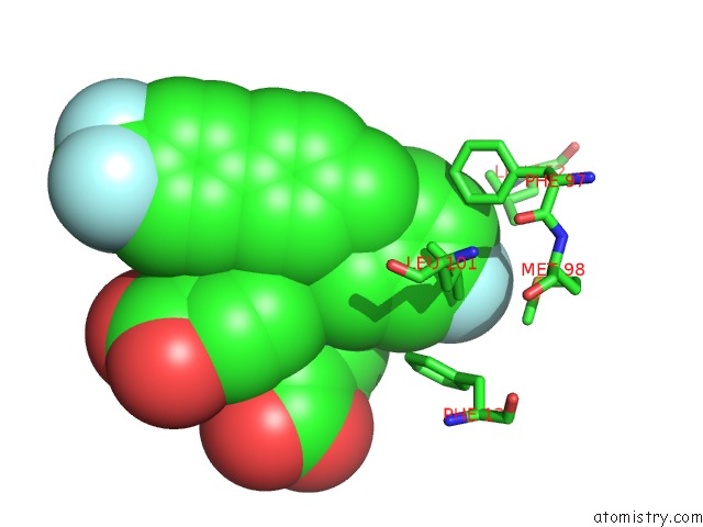
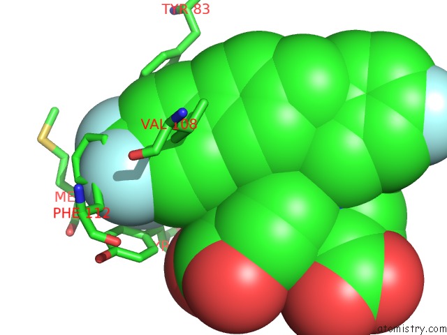
Fluorine binding site 3 out
of 8 in 4iah
Go back to
Fluorine Binding Sites List in 4iah
Fluorine binding site 3 out
of 8 in the Crystal Structure of Bay 60-2770 Bound C139A H-Nox Domain with S- Nitrosylated Conserved C122
 Mono view
 Stereo pair view
|
A full contact list of Fluorine with other atoms in the F binding
site number 3 of Crystal Structure of Bay 60-2770 Bound C139A H-Nox Domain with S- Nitrosylated Conserved C122 within 5.0Å range:
probe
|
atom
|
residue
|
distance (Å)
|
B
|
Occ
|
A:F201
b:33.4
occ:1.00
|
FAJ
|
A:1DX201
|
0.0
|
33.4
|
1.0
|
CAI
|
A:1DX201
|
1.3
|
33.6
|
1.0
|
FAE
|
A:1DX201
|
2.2
|
33.8
|
1.0
|
FAK
|
A:1DX201
|
2.2
|
33.9
|
1.0
|
CAH
|
A:1DX201
|
2.3
|
33.1
|
1.0
|
CAG
|
A:1DX201
|
3.1
|
32.7
|
1.0
|
O
|
A:GLY39
|
3.2
|
39.3
|
1.0
|
CZD
|
A:1DX201
|
3.2
|
32.7
|
1.0
|
CB
|
A:TYR2
|
3.7
|
34.7
|
1.0
|
CB
|
A:LEU4
|
3.8
|
33.4
|
1.0
|
CA
|
A:MET40
|
4.0
|
38.9
|
1.0
|
C
|
A:GLY39
|
4.0
|
39.3
|
1.0
|
CG
|
A:TYR2
|
4.0
|
35.2
|
1.0
|
CD2
|
A:LEU4
|
4.2
|
33.4
|
1.0
|
OH
|
A:TYR83
|
4.2
|
32.9
|
1.0
|
CG
|
A:MET40
|
4.3
|
38.8
|
1.0
|
CAJ
|
A:1DX201
|
4.3
|
32.3
|
1.0
|
CD2
|
A:TYR2
|
4.4
|
35.7
|
1.0
|
N
|
A:MET40
|
4.4
|
39.1
|
1.0
|
CAK
|
A:1DX201
|
4.4
|
32.4
|
1.0
|
CE1
|
A:PHE112
|
4.5
|
44.0
|
1.0
|
CB
|
A:MET40
|
4.6
|
38.9
|
1.0
|
CZ
|
A:PHE112
|
4.6
|
44.1
|
1.0
|
CG
|
A:LEU4
|
4.6
|
33.3
|
1.0
|
CD1
|
A:TYR2
|
4.6
|
35.6
|
1.0
|
CD1
|
A:PHE112
|
4.6
|
43.8
|
1.0
|
CE2
|
A:PHE112
|
4.7
|
44.0
|
1.0
|
CG
|
A:PHE112
|
4.8
|
43.3
|
1.0
|
N
|
A:LEU4
|
4.8
|
33.5
|
1.0
|
CD2
|
A:PHE112
|
4.9
|
43.7
|
1.0
|
CBI
|
A:1DX201
|
4.9
|
31.9
|
1.0
|
CA
|
A:LEU4
|
4.9
|
33.4
|
1.0
|
C
|
A:MET40
|
5.0
|
38.9
|
1.0
|
|
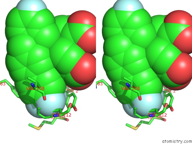
Fluorine binding site 4 out
of 8 in 4iah
Go back to
Fluorine Binding Sites List in 4iah
Fluorine binding site 4 out
of 8 in the Crystal Structure of Bay 60-2770 Bound C139A H-Nox Domain with S- Nitrosylated Conserved C122
 Mono view
 Stereo pair view
|
A full contact list of Fluorine with other atoms in the F binding
site number 4 of Crystal Structure of Bay 60-2770 Bound C139A H-Nox Domain with S- Nitrosylated Conserved C122 within 5.0Å range:
probe
|
atom
|
residue
|
distance (Å)
|
B
|
Occ
|
A:F201
b:33.9
occ:1.00
|
FAK
|
A:1DX201
|
0.0
|
33.9
|
1.0
|
CAI
|
A:1DX201
|
1.3
|
33.6
|
1.0
|
FAE
|
A:1DX201
|
2.2
|
33.8
|
1.0
|
FAJ
|
A:1DX201
|
2.2
|
33.4
|
1.0
|
CAH
|
A:1DX201
|
2.3
|
33.1
|
1.0
|
CZD
|
A:1DX201
|
2.7
|
32.7
|
1.0
|
CG
|
A:PHE112
|
3.0
|
43.3
|
1.0
|
CD2
|
A:PHE112
|
3.2
|
43.7
|
1.0
|
CD1
|
A:TYR2
|
3.3
|
35.6
|
1.0
|
CG
|
A:TYR2
|
3.3
|
35.2
|
1.0
|
CD1
|
A:PHE112
|
3.4
|
43.8
|
1.0
|
CB
|
A:PHE112
|
3.6
|
43.0
|
1.0
|
CAG
|
A:1DX201
|
3.6
|
32.7
|
1.0
|
CE2
|
A:PHE112
|
3.7
|
44.0
|
1.0
|
CE1
|
A:TYR2
|
3.7
|
35.9
|
1.0
|
CD2
|
A:TYR2
|
3.8
|
35.7
|
1.0
|
CB
|
A:TYR2
|
3.8
|
34.7
|
1.0
|
CE1
|
A:PHE112
|
3.8
|
44.0
|
1.0
|
CZ
|
A:PHE112
|
3.9
|
44.1
|
1.0
|
CAK
|
A:1DX201
|
4.0
|
32.4
|
1.0
|
CZ
|
A:TYR2
|
4.1
|
36.0
|
1.0
|
CE2
|
A:TYR2
|
4.2
|
35.9
|
1.0
|
CAJ
|
A:1DX201
|
4.7
|
32.3
|
1.0
|
O
|
A:GLY39
|
4.8
|
39.3
|
1.0
|
CBI
|
A:1DX201
|
4.9
|
31.9
|
1.0
|
CA
|
A:MET40
|
4.9
|
38.9
|
1.0
|
CD1
|
A:LEU115
|
5.0
|
39.3
|
1.0
|
CG1
|
A:VAL108
|
5.0
|
40.3
|
1.0
|
|
Fluorine binding site 5 out
of 8 in 4iah
Go back to
Fluorine Binding Sites List in 4iah
Fluorine binding site 5 out
of 8 in the Crystal Structure of Bay 60-2770 Bound C139A H-Nox Domain with S- Nitrosylated Conserved C122
 Mono view
 Stereo pair view
|
A full contact list of Fluorine with other atoms in the F binding
site number 5 of Crystal Structure of Bay 60-2770 Bound C139A H-Nox Domain with S- Nitrosylated Conserved C122 within 5.0Å range:
probe
|
atom
|
residue
|
distance (Å)
|
B
|
Occ
|
B:F201
b:35.0
occ:1.00
|
FAA
|
B:1DX201
|
0.0
|
35.0
|
1.0
|
CAC
|
B:1DX201
|
1.3
|
34.8
|
1.0
|
CAB
|
B:1DX201
|
2.4
|
34.8
|
1.0
|
CAD
|
B:1DX201
|
2.4
|
34.5
|
1.0
|
CB
|
B:LEU148
|
3.6
|
25.3
|
1.0
|
CBN
|
B:1DX201
|
3.6
|
34.2
|
1.0
|
CAE
|
B:1DX201
|
3.6
|
34.7
|
1.0
|
CD1
|
B:LEU101
|
3.7
|
31.7
|
1.0
|
CD1
|
B:LEU148
|
3.8
|
25.0
|
1.0
|
CD1
|
B:LEU152
|
4.1
|
28.2
|
1.0
|
CBO
|
B:1DX201
|
4.1
|
34.7
|
1.0
|
CG
|
B:LEU148
|
4.2
|
25.2
|
1.0
|
CE
|
B:MET98
|
4.4
|
29.0
|
1.0
|
SD
|
B:MET98
|
4.4
|
29.4
|
1.0
|
CD2
|
B:LEU148
|
4.5
|
25.1
|
1.0
|
CZ
|
B:PHE120
|
4.6
|
33.3
|
1.0
|
CD2
|
B:LEU101
|
4.6
|
31.9
|
1.0
|
C
|
B:LEU148
|
4.7
|
25.4
|
1.0
|
CE2
|
B:PHE97
|
4.7
|
28.2
|
1.0
|
CA
|
B:LEU148
|
4.7
|
25.3
|
1.0
|
CG
|
B:LEU101
|
4.7
|
31.8
|
1.0
|
CG
|
B:LEU152
|
4.8
|
28.4
|
1.0
|
O
|
B:LEU148
|
4.8
|
25.4
|
1.0
|
CE1
|
B:PHE120
|
4.9
|
33.4
|
1.0
|
CBA
|
B:1DX201
|
4.9
|
33.6
|
1.0
|
CE2
|
B:PHE120
|
5.0
|
33.5
|
1.0
|
|
Fluorine binding site 6 out
of 8 in 4iah
Go back to
Fluorine Binding Sites List in 4iah
Fluorine binding site 6 out
of 8 in the Crystal Structure of Bay 60-2770 Bound C139A H-Nox Domain with S- Nitrosylated Conserved C122
 Mono view
 Stereo pair view
|
A full contact list of Fluorine with other atoms in the F binding
site number 6 of Crystal Structure of Bay 60-2770 Bound C139A H-Nox Domain with S- Nitrosylated Conserved C122 within 5.0Å range:
probe
|
atom
|
residue
|
distance (Å)
|
B
|
Occ
|
B:F201
b:37.6
occ:1.00
|
FAE
|
B:1DX201
|
0.0
|
37.6
|
1.0
|
CAI
|
B:1DX201
|
1.3
|
37.7
|
1.0
|
FAK
|
B:1DX201
|
2.2
|
37.7
|
1.0
|
FAJ
|
B:1DX201
|
2.2
|
38.0
|
1.0
|
CAH
|
B:1DX201
|
2.3
|
37.6
|
1.0
|
CZD
|
B:1DX201
|
2.9
|
37.8
|
1.0
|
CB
|
B:TYR2
|
3.1
|
34.5
|
1.0
|
CD2
|
B:TYR2
|
3.2
|
35.3
|
1.0
|
O
|
B:GLY39
|
3.2
|
41.6
|
1.0
|
CG
|
B:TYR2
|
3.3
|
35.0
|
1.0
|
CAG
|
B:1DX201
|
3.3
|
37.5
|
1.0
|
CB
|
B:LEU4
|
4.0
|
33.8
|
1.0
|
C
|
B:GLY39
|
4.1
|
41.6
|
1.0
|
CE2
|
B:TYR2
|
4.1
|
35.4
|
1.0
|
CA
|
B:MET40
|
4.2
|
41.3
|
1.0
|
CAK
|
B:1DX201
|
4.2
|
37.5
|
1.0
|
CD1
|
B:TYR2
|
4.3
|
35.3
|
1.0
|
CD2
|
B:LEU4
|
4.4
|
33.2
|
1.0
|
CAJ
|
B:1DX201
|
4.5
|
37.3
|
1.0
|
CA
|
B:TYR2
|
4.5
|
34.4
|
1.0
|
CE1
|
B:PHE112
|
4.5
|
42.3
|
1.0
|
CD1
|
B:PHE112
|
4.5
|
42.0
|
1.0
|
N
|
B:MET40
|
4.6
|
41.5
|
1.0
|
CZ
|
B:PHE112
|
4.6
|
42.5
|
1.0
|
CG
|
B:PHE112
|
4.7
|
41.6
|
1.0
|
OH
|
B:TYR83
|
4.7
|
27.3
|
1.0
|
CE2
|
B:PHE112
|
4.7
|
42.5
|
1.0
|
CD2
|
B:PHE112
|
4.8
|
42.0
|
1.0
|
N
|
B:LEU4
|
4.8
|
34.0
|
1.0
|
CG
|
B:MET40
|
4.8
|
41.5
|
1.0
|
C
|
B:TYR2
|
4.8
|
34.3
|
1.0
|
CG
|
B:LEU4
|
4.8
|
33.5
|
1.0
|
CBI
|
B:1DX201
|
4.9
|
37.2
|
1.0
|
CB
|
B:MET40
|
5.0
|
41.3
|
1.0
|
CZ
|
B:TYR2
|
5.0
|
35.7
|
1.0
|
C
|
B:MET40
|
5.0
|
41.2
|
1.0
|
|
Fluorine binding site 7 out
of 8 in 4iah
Go back to
Fluorine Binding Sites List in 4iah
Fluorine binding site 7 out
of 8 in the Crystal Structure of Bay 60-2770 Bound C139A H-Nox Domain with S- Nitrosylated Conserved C122
 Mono view
 Stereo pair view
|
A full contact list of Fluorine with other atoms in the F binding
site number 7 of Crystal Structure of Bay 60-2770 Bound C139A H-Nox Domain with S- Nitrosylated Conserved C122 within 5.0Å range:
probe
|
atom
|
residue
|
distance (Å)
|
B
|
Occ
|
B:F201
b:38.0
occ:1.00
|
FAJ
|
B:1DX201
|
0.0
|
38.0
|
1.0
|
CAI
|
B:1DX201
|
1.3
|
37.7
|
1.0
|
FAK
|
B:1DX201
|
2.2
|
37.7
|
1.0
|
FAE
|
B:1DX201
|
2.2
|
37.6
|
1.0
|
CAH
|
B:1DX201
|
2.3
|
37.6
|
1.0
|
CD2
|
B:PHE112
|
2.6
|
42.0
|
1.0
|
CG
|
B:PHE112
|
2.7
|
41.6
|
1.0
|
CE2
|
B:PHE112
|
2.8
|
42.5
|
1.0
|
CZD
|
B:1DX201
|
2.9
|
37.8
|
1.0
|
CD1
|
B:PHE112
|
3.0
|
42.0
|
1.0
|
CZ
|
B:PHE112
|
3.1
|
42.5
|
1.0
|
CE1
|
B:PHE112
|
3.2
|
42.3
|
1.0
|
CAG
|
B:1DX201
|
3.4
|
37.5
|
1.0
|
CB
|
B:PHE112
|
3.5
|
41.3
|
1.0
|
CD2
|
B:TYR2
|
3.5
|
35.3
|
1.0
|
CE2
|
B:TYR2
|
3.8
|
35.4
|
1.0
|
CG
|
B:TYR2
|
4.0
|
35.0
|
1.0
|
CAK
|
B:1DX201
|
4.2
|
37.5
|
1.0
|
CZ
|
B:TYR2
|
4.4
|
35.7
|
1.0
|
CB
|
B:TYR2
|
4.5
|
34.5
|
1.0
|
CD1
|
B:TYR2
|
4.5
|
35.3
|
1.0
|
OH
|
B:TYR83
|
4.6
|
27.3
|
1.0
|
CG1
|
B:VAL108
|
4.6
|
38.8
|
1.0
|
CAJ
|
B:1DX201
|
4.6
|
37.3
|
1.0
|
CE1
|
B:TYR2
|
4.7
|
35.6
|
1.0
|
CA
|
B:MET40
|
4.9
|
41.3
|
1.0
|
CBI
|
B:1DX201
|
4.9
|
37.2
|
1.0
|
CG
|
B:MET40
|
5.0
|
41.5
|
1.0
|
CA
|
B:PHE112
|
5.0
|
41.2
|
1.0
|
|
Fluorine binding site 8 out
of 8 in 4iah
Go back to
Fluorine Binding Sites List in 4iah
Fluorine binding site 8 out
of 8 in the Crystal Structure of Bay 60-2770 Bound C139A H-Nox Domain with S- Nitrosylated Conserved C122
 Mono view
 Stereo pair view
|
A full contact list of Fluorine with other atoms in the F binding
site number 8 of Crystal Structure of Bay 60-2770 Bound C139A H-Nox Domain with S- Nitrosylated Conserved C122 within 5.0Å range:
probe
|
atom
|
residue
|
distance (Å)
|
B
|
Occ
|
B:F201
b:37.7
occ:1.00
|
FAK
|
B:1DX201
|
0.0
|
37.7
|
1.0
|
CAI
|
B:1DX201
|
1.3
|
37.7
|
1.0
|
FAE
|
B:1DX201
|
2.2
|
37.6
|
1.0
|
FAJ
|
B:1DX201
|
2.2
|
38.0
|
1.0
|
CAH
|
B:1DX201
|
2.3
|
37.6
|
1.0
|
CAG
|
B:1DX201
|
2.7
|
37.5
|
1.0
|
OH
|
B:TYR83
|
2.7
|
27.3
|
1.0
|
CZ
|
B:PHE112
|
3.0
|
42.5
|
1.0
|
CE1
|
B:PHE112
|
3.3
|
42.3
|
1.0
|
CE2
|
B:PHE112
|
3.3
|
42.5
|
1.0
|
CZD
|
B:1DX201
|
3.6
|
37.8
|
1.0
|
CZ
|
B:TYR83
|
3.7
|
28.1
|
1.0
|
CG
|
B:MET40
|
3.9
|
41.5
|
1.0
|
CD1
|
B:PHE112
|
3.9
|
42.0
|
1.0
|
CD2
|
B:PHE112
|
4.0
|
42.0
|
1.0
|
CAJ
|
B:1DX201
|
4.0
|
37.3
|
1.0
|
CG
|
B:PHE112
|
4.2
|
41.6
|
1.0
|
O
|
B:GLY39
|
4.4
|
41.6
|
1.0
|
CD2
|
B:LEU4
|
4.4
|
33.2
|
1.0
|
CE2
|
B:TYR83
|
4.4
|
28.3
|
1.0
|
CE1
|
B:TYR83
|
4.4
|
28.2
|
1.0
|
CA
|
B:MET40
|
4.5
|
41.3
|
1.0
|
CB
|
B:MET40
|
4.6
|
41.3
|
1.0
|
CD2
|
B:TYR2
|
4.7
|
35.3
|
1.0
|
CAK
|
B:1DX201
|
4.7
|
37.5
|
1.0
|
CE
|
B:MET40
|
4.7
|
41.9
|
1.0
|
C
|
B:GLY39
|
4.8
|
41.6
|
1.0
|
CB
|
B:LEU4
|
4.9
|
33.8
|
1.0
|
N
|
B:MET40
|
4.9
|
41.5
|
1.0
|
CBI
|
B:1DX201
|
4.9
|
37.2
|
1.0
|
SD
|
B:MET40
|
5.0
|
41.9
|
1.0
|
|
Reference:
V.Kumar,
F.Martin,
M.G.Hahn,
M.Schaefer,
J.S.Stamler,
J.P.Stasch,
F.Van Den Akker.
Insights Into Bay 60-2770 Activation and S-Nitrosylation-Dependent Desensitization of Soluble Guanylyl Cyclase Via Crystal Structures of Homologous Nostoc H-Nox Domain Complexes. Biochemistry V. 52 3601 2013.
ISSN: ISSN 0006-2960
PubMed: 23614626
DOI: 10.1021/BI301657W
Page generated: Thu Aug 1 02:19:56 2024
|