Fluorine »
PDB 4iju-4j0t »
4iwf »
Fluorine in PDB 4iwf: Crystal Structure of the Estrogen Receptor Alpha Ligand-Binding Domain in Complex with A Dynamic Oxime-Derivative
Protein crystallography data
The structure of Crystal Structure of the Estrogen Receptor Alpha Ligand-Binding Domain in Complex with A Dynamic Oxime-Derivative, PDB code: 4iwf
was solved by
J.C.Nwachukwu,
S.Srinivasan,
A.A.Parent,
V.Cavett,
J.Nowak,
T.S.Hughes,
D.J.Kojetin,
J.A.Katzenellenbogen,
K.W.Nettles,
with X-Ray Crystallography technique. A brief refinement statistics is given in the table below:
| Resolution Low / High (Å) | 40.42 / 1.93 |
| Space group | P 1 21 1 |
| Cell size a, b, c (Å), α, β, γ (°) | 55.897, 83.036, 58.191, 90.00, 108.54, 90.00 |
| R / Rfree (%) | 17.3 / 20.9 |
Other elements in 4iwf:
The structure of Crystal Structure of the Estrogen Receptor Alpha Ligand-Binding Domain in Complex with A Dynamic Oxime-Derivative also contains other interesting chemical elements:
| Chlorine | (Cl) | 3 atoms |
Fluorine Binding Sites:
The binding sites of Fluorine atom in the Crystal Structure of the Estrogen Receptor Alpha Ligand-Binding Domain in Complex with A Dynamic Oxime-Derivative
(pdb code 4iwf). This binding sites where shown within
5.0 Angstroms radius around Fluorine atom.
In total 3 binding sites of Fluorine where determined in the Crystal Structure of the Estrogen Receptor Alpha Ligand-Binding Domain in Complex with A Dynamic Oxime-Derivative, PDB code: 4iwf:
Jump to Fluorine binding site number: 1; 2; 3;
In total 3 binding sites of Fluorine where determined in the Crystal Structure of the Estrogen Receptor Alpha Ligand-Binding Domain in Complex with A Dynamic Oxime-Derivative, PDB code: 4iwf:
Jump to Fluorine binding site number: 1; 2; 3;
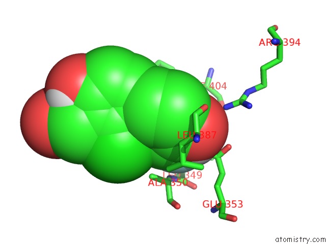
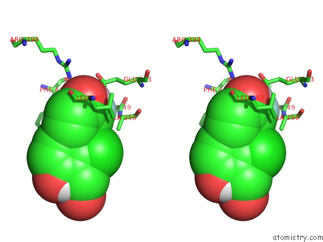
Fluorine binding site 1 out of 3 in 4iwf
Go back to
Fluorine binding site 1 out
of 3 in the Crystal Structure of the Estrogen Receptor Alpha Ligand-Binding Domain in Complex with A Dynamic Oxime-Derivative
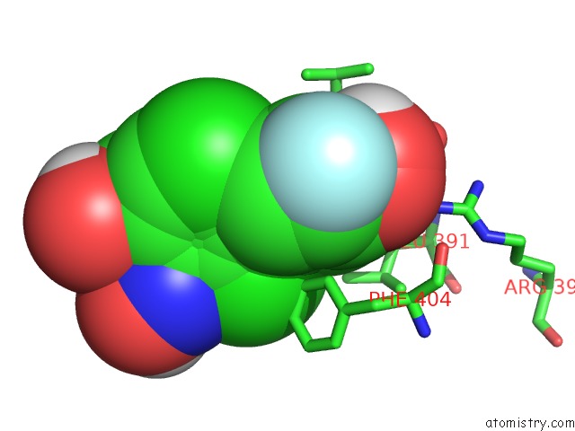
Mono view
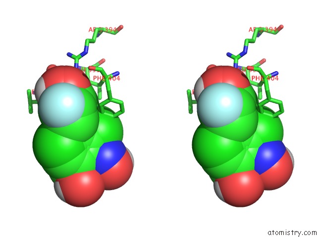
Stereo pair view
Mono view
Stereo pair view
A full contact list of Fluorine with other atoms in the F binding
site number 1 of Crystal Structure of the Estrogen Receptor Alpha Ligand-Binding Domain in Complex with A Dynamic Oxime-Derivative within 5.0Å range:
|
Fluorine binding site 2 out of 3 in 4iwf
Go back to
Fluorine binding site 2 out
of 3 in the Crystal Structure of the Estrogen Receptor Alpha Ligand-Binding Domain in Complex with A Dynamic Oxime-Derivative
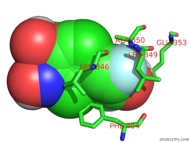
Mono view
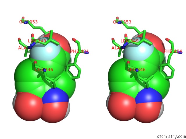
Stereo pair view
Mono view
Stereo pair view
A full contact list of Fluorine with other atoms in the F binding
site number 2 of Crystal Structure of the Estrogen Receptor Alpha Ligand-Binding Domain in Complex with A Dynamic Oxime-Derivative within 5.0Å range:
|
Fluorine binding site 3 out of 3 in 4iwf
Go back to
Fluorine binding site 3 out
of 3 in the Crystal Structure of the Estrogen Receptor Alpha Ligand-Binding Domain in Complex with A Dynamic Oxime-Derivative
Mono view
Stereo pair view
Mono view
Stereo pair view
A full contact list of Fluorine with other atoms in the F binding
site number 3 of Crystal Structure of the Estrogen Receptor Alpha Ligand-Binding Domain in Complex with A Dynamic Oxime-Derivative within 5.0Å range:
|
Reference:
S.Srinivasan,
J.C.Nwachukwu,
A.A.Parent,
V.Cavett,
J.Nowak,
T.S.Hughes,
D.J.Kojetin,
J.A.Katzenellenbogen,
K.W.Nettles.
Ligand Binding Dynamics Rewire Cellular Signaling Via Estrogen Receptor-Alpha Nat.Chem.Biol. V. 9 326 2013.
ISSN: ISSN 1552-4450
PubMed: 23524984
DOI: 10.1038/NCHEMBIO.1214
Page generated: Thu Aug 1 02:39:58 2024
ISSN: ISSN 1552-4450
PubMed: 23524984
DOI: 10.1038/NCHEMBIO.1214
Last articles
Zn in 9MJ5Zn in 9HNW
Zn in 9G0L
Zn in 9FNE
Zn in 9DZN
Zn in 9E0I
Zn in 9D32
Zn in 9DAK
Zn in 8ZXC
Zn in 8ZUF