Fluorine »
PDB 4lvt-4mm8 »
4lxi »
Fluorine in PDB 4lxi: Crystal Structure of the S105A Mutant of A Carbon-Carbon Bond Hydrolase, DXNB2 From Sphingomonas Wittichii RW1, in Complex with 5, 8-Dif Hopda
Enzymatic activity of Crystal Structure of the S105A Mutant of A Carbon-Carbon Bond Hydrolase, DXNB2 From Sphingomonas Wittichii RW1, in Complex with 5, 8-Dif Hopda
All present enzymatic activity of Crystal Structure of the S105A Mutant of A Carbon-Carbon Bond Hydrolase, DXNB2 From Sphingomonas Wittichii RW1, in Complex with 5, 8-Dif Hopda:
3.7.1.8;
3.7.1.8;
Protein crystallography data
The structure of Crystal Structure of the S105A Mutant of A Carbon-Carbon Bond Hydrolase, DXNB2 From Sphingomonas Wittichii RW1, in Complex with 5, 8-Dif Hopda, PDB code: 4lxi
was solved by
S.Bhowmik,
J.T.Bolin,
with X-Ray Crystallography technique. A brief refinement statistics is given in the table below:
| Resolution Low / High (Å) | 57.26 / 2.17 |
| Space group | P 65 2 2 |
| Cell size a, b, c (Å), α, β, γ (°) | 66.155, 66.155, 342.180, 90.00, 90.00, 120.00 |
| R / Rfree (%) | 20.4 / 25.8 |
Other elements in 4lxi:
The structure of Crystal Structure of the S105A Mutant of A Carbon-Carbon Bond Hydrolase, DXNB2 From Sphingomonas Wittichii RW1, in Complex with 5, 8-Dif Hopda also contains other interesting chemical elements:
| Sodium | (Na) | 1 atom |
Fluorine Binding Sites:
The binding sites of Fluorine atom in the Crystal Structure of the S105A Mutant of A Carbon-Carbon Bond Hydrolase, DXNB2 From Sphingomonas Wittichii RW1, in Complex with 5, 8-Dif Hopda
(pdb code 4lxi). This binding sites where shown within
5.0 Angstroms radius around Fluorine atom.
In total 2 binding sites of Fluorine where determined in the Crystal Structure of the S105A Mutant of A Carbon-Carbon Bond Hydrolase, DXNB2 From Sphingomonas Wittichii RW1, in Complex with 5, 8-Dif Hopda, PDB code: 4lxi:
Jump to Fluorine binding site number: 1; 2;
In total 2 binding sites of Fluorine where determined in the Crystal Structure of the S105A Mutant of A Carbon-Carbon Bond Hydrolase, DXNB2 From Sphingomonas Wittichii RW1, in Complex with 5, 8-Dif Hopda, PDB code: 4lxi:
Jump to Fluorine binding site number: 1; 2;
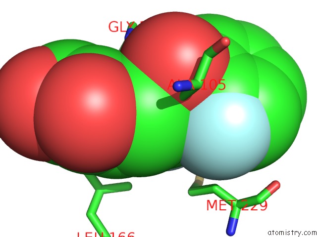
Fluorine binding site 1 out of 2 in 4lxi
Go back to
Fluorine binding site 1 out
of 2 in the Crystal Structure of the S105A Mutant of A Carbon-Carbon Bond Hydrolase, DXNB2 From Sphingomonas Wittichii RW1, in Complex with 5, 8-Dif Hopda
Mono view

Stereo pair view
Mono view
Stereo pair view
A full contact list of Fluorine with other atoms in the F binding
site number 1 of Crystal Structure of the S105A Mutant of A Carbon-Carbon Bond Hydrolase, DXNB2 From Sphingomonas Wittichii RW1, in Complex with 5, 8-Dif Hopda within 5.0Å range:
|
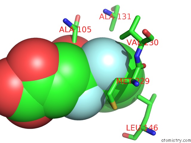
Fluorine binding site 2 out of 2 in 4lxi
Go back to
Fluorine binding site 2 out
of 2 in the Crystal Structure of the S105A Mutant of A Carbon-Carbon Bond Hydrolase, DXNB2 From Sphingomonas Wittichii RW1, in Complex with 5, 8-Dif Hopda
Mono view

Stereo pair view
Mono view
Stereo pair view
A full contact list of Fluorine with other atoms in the F binding
site number 2 of Crystal Structure of the S105A Mutant of A Carbon-Carbon Bond Hydrolase, DXNB2 From Sphingomonas Wittichii RW1, in Complex with 5, 8-Dif Hopda within 5.0Å range:
|
Reference:
A.C.Ruzzini,
S.Bhowmik,
S.Ghosh,
K.C.Yam,
J.T.Bolin,
L.D.Eltis.
A Substrate-Assisted Mechanism of Nucleophile Activation in A Ser-His-Asp Containing C-C Bond Hydrolase. Biochemistry V. 52 7428 2013.
ISSN: ISSN 0006-2960
PubMed: 24067021
DOI: 10.1021/BI401156A
Page generated: Thu Aug 1 03:32:17 2024
ISSN: ISSN 0006-2960
PubMed: 24067021
DOI: 10.1021/BI401156A
Last articles
Ca in 5N3YCa in 5N31
Ca in 5N3V
Ca in 5N34
Ca in 5N2Z
Ca in 5N2X
Ca in 5N2T
Ca in 5N1B
Ca in 5MWF
Ca in 5N2J