Fluorine »
PDB 4mm9-4ncg »
4muw »
Fluorine in PDB 4muw: Crystal Structure of PDE10A with Novel Keto-Benzimidazole Inhibitor
Enzymatic activity of Crystal Structure of PDE10A with Novel Keto-Benzimidazole Inhibitor
All present enzymatic activity of Crystal Structure of PDE10A with Novel Keto-Benzimidazole Inhibitor:
3.1.4.17; 3.1.4.35;
3.1.4.17; 3.1.4.35;
Protein crystallography data
The structure of Crystal Structure of PDE10A with Novel Keto-Benzimidazole Inhibitor, PDB code: 4muw
was solved by
S.Chmait,
S.Jordan,
with X-Ray Crystallography technique. A brief refinement statistics is given in the table below:
| Resolution Low / High (Å) | 29.19 / 2.64 |
| Space group | F 2 3 |
| Cell size a, b, c (Å), α, β, γ (°) | 252.659, 252.659, 252.659, 90.00, 90.00, 90.00 |
| R / Rfree (%) | 17.1 / 19.6 |
Other elements in 4muw:
The structure of Crystal Structure of PDE10A with Novel Keto-Benzimidazole Inhibitor also contains other interesting chemical elements:
| Zinc | (Zn) | 4 atoms |
Fluorine Binding Sites:
The binding sites of Fluorine atom in the Crystal Structure of PDE10A with Novel Keto-Benzimidazole Inhibitor
(pdb code 4muw). This binding sites where shown within
5.0 Angstroms radius around Fluorine atom.
In total 4 binding sites of Fluorine where determined in the Crystal Structure of PDE10A with Novel Keto-Benzimidazole Inhibitor, PDB code: 4muw:
Jump to Fluorine binding site number: 1; 2; 3; 4;
In total 4 binding sites of Fluorine where determined in the Crystal Structure of PDE10A with Novel Keto-Benzimidazole Inhibitor, PDB code: 4muw:
Jump to Fluorine binding site number: 1; 2; 3; 4;
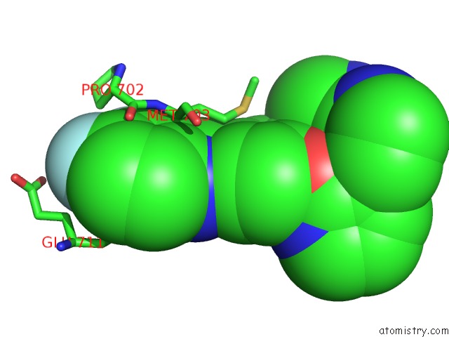
Fluorine binding site 1 out of 4 in 4muw
Go back to
Fluorine binding site 1 out
of 4 in the Crystal Structure of PDE10A with Novel Keto-Benzimidazole Inhibitor

Mono view
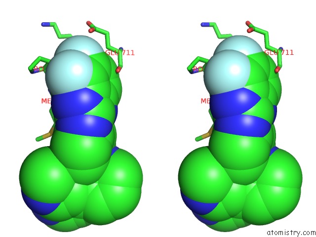
Stereo pair view
Mono view
Stereo pair view
A full contact list of Fluorine with other atoms in the F binding
site number 1 of Crystal Structure of PDE10A with Novel Keto-Benzimidazole Inhibitor within 5.0Å range:
|
Fluorine binding site 2 out of 4 in 4muw
Go back to
Fluorine binding site 2 out
of 4 in the Crystal Structure of PDE10A with Novel Keto-Benzimidazole Inhibitor
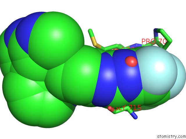
Mono view

Stereo pair view
Mono view
Stereo pair view
A full contact list of Fluorine with other atoms in the F binding
site number 2 of Crystal Structure of PDE10A with Novel Keto-Benzimidazole Inhibitor within 5.0Å range:
|
Fluorine binding site 3 out of 4 in 4muw
Go back to
Fluorine binding site 3 out
of 4 in the Crystal Structure of PDE10A with Novel Keto-Benzimidazole Inhibitor
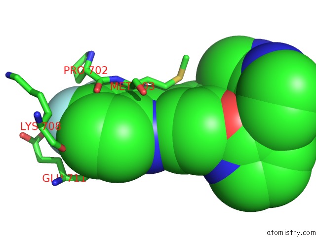
Mono view
Stereo pair view
Mono view
Stereo pair view
A full contact list of Fluorine with other atoms in the F binding
site number 3 of Crystal Structure of PDE10A with Novel Keto-Benzimidazole Inhibitor within 5.0Å range:
|
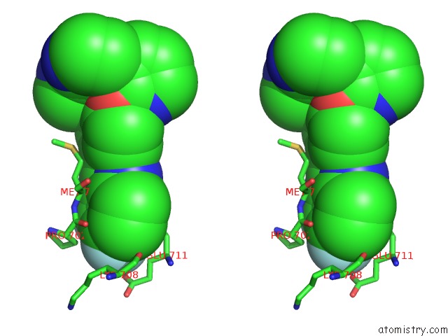
Fluorine binding site 4 out of 4 in 4muw
Go back to
Fluorine binding site 4 out
of 4 in the Crystal Structure of PDE10A with Novel Keto-Benzimidazole Inhibitor
Mono view

Stereo pair view
Mono view
Stereo pair view
A full contact list of Fluorine with other atoms in the F binding
site number 4 of Crystal Structure of PDE10A with Novel Keto-Benzimidazole Inhibitor within 5.0Å range:
|
Reference:
E.Hu,
R.K.Kunz,
N.Chen,
S.Rumfelt,
A.Siegmund,
K.Andrews,
S.Chmait,
S.Zhao,
C.Davis,
H.Chen,
D.Lester-Zeiner,
J.Ma,
C.Biorn,
J.Shi,
A.Porter,
J.Treanor,
J.R.Allen.
Design, Optimization, and Biological Evaluation of Novel Keto-Benzimidazoles As Potent and Selective Inhibitors of Phosphodiesterase 10A (PDE10A). J.Med.Chem. V. 56 8781 2013.
ISSN: ISSN 0022-2623
PubMed: 24102193
DOI: 10.1021/JM401234W
Page generated: Thu Aug 1 04:02:17 2024
ISSN: ISSN 0022-2623
PubMed: 24102193
DOI: 10.1021/JM401234W
Last articles
Zn in 9MJ5Zn in 9HNW
Zn in 9G0L
Zn in 9FNE
Zn in 9DZN
Zn in 9E0I
Zn in 9D32
Zn in 9DAK
Zn in 8ZXC
Zn in 8ZUF