Fluorine »
PDB 4mm9-4ncg »
4n07 »
Fluorine in PDB 4n07: Crystal Structure of the GLUA2 Ligand-Binding Domain (S1S2J-L483Y- N754S) in Complex with Glutamate and Bpam-344 at 1.87 A Resolution
Protein crystallography data
The structure of Crystal Structure of the GLUA2 Ligand-Binding Domain (S1S2J-L483Y- N754S) in Complex with Glutamate and Bpam-344 at 1.87 A Resolution, PDB code: 4n07
was solved by
A.B.Noerholm,
K.Frydenvang,
J.S.Kastrup,
with X-Ray Crystallography technique. A brief refinement statistics is given in the table below:
| Resolution Low / High (Å) | 28.48 / 1.87 |
| Space group | P 21 21 2 |
| Cell size a, b, c (Å), α, β, γ (°) | 113.904, 164.034, 47.284, 90.00, 90.00, 90.00 |
| R / Rfree (%) | 17.5 / 21.1 |
Other elements in 4n07:
The structure of Crystal Structure of the GLUA2 Ligand-Binding Domain (S1S2J-L483Y- N754S) in Complex with Glutamate and Bpam-344 at 1.87 A Resolution also contains other interesting chemical elements:
| Arsenic | (As) | 1 atom |
| Zinc | (Zn) | 10 atoms |
Fluorine Binding Sites:
The binding sites of Fluorine atom in the Crystal Structure of the GLUA2 Ligand-Binding Domain (S1S2J-L483Y- N754S) in Complex with Glutamate and Bpam-344 at 1.87 A Resolution
(pdb code 4n07). This binding sites where shown within
5.0 Angstroms radius around Fluorine atom.
In total 3 binding sites of Fluorine where determined in the Crystal Structure of the GLUA2 Ligand-Binding Domain (S1S2J-L483Y- N754S) in Complex with Glutamate and Bpam-344 at 1.87 A Resolution, PDB code: 4n07:
Jump to Fluorine binding site number: 1; 2; 3;
In total 3 binding sites of Fluorine where determined in the Crystal Structure of the GLUA2 Ligand-Binding Domain (S1S2J-L483Y- N754S) in Complex with Glutamate and Bpam-344 at 1.87 A Resolution, PDB code: 4n07:
Jump to Fluorine binding site number: 1; 2; 3;
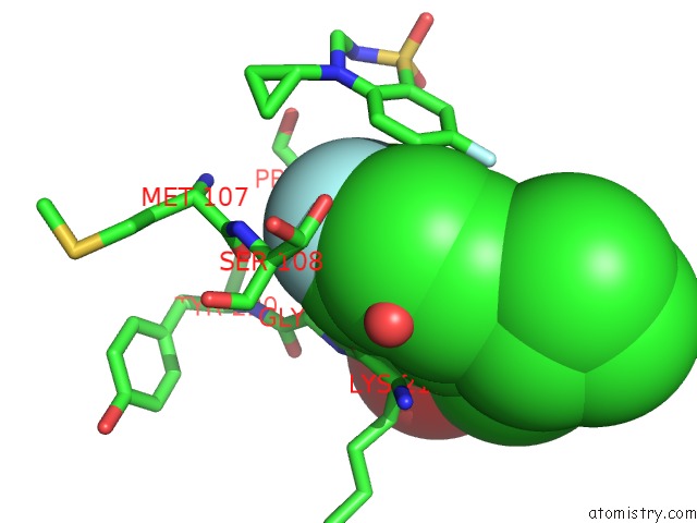
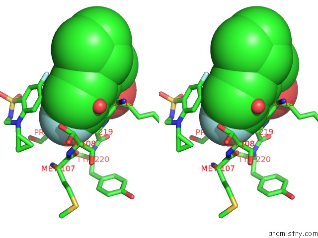
Fluorine binding site 1 out of 3 in 4n07
Go back to
Fluorine binding site 1 out
of 3 in the Crystal Structure of the GLUA2 Ligand-Binding Domain (S1S2J-L483Y- N754S) in Complex with Glutamate and Bpam-344 at 1.87 A Resolution
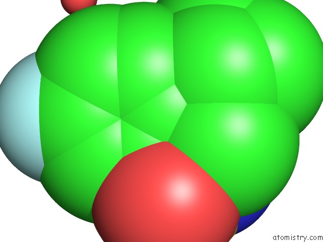
Mono view
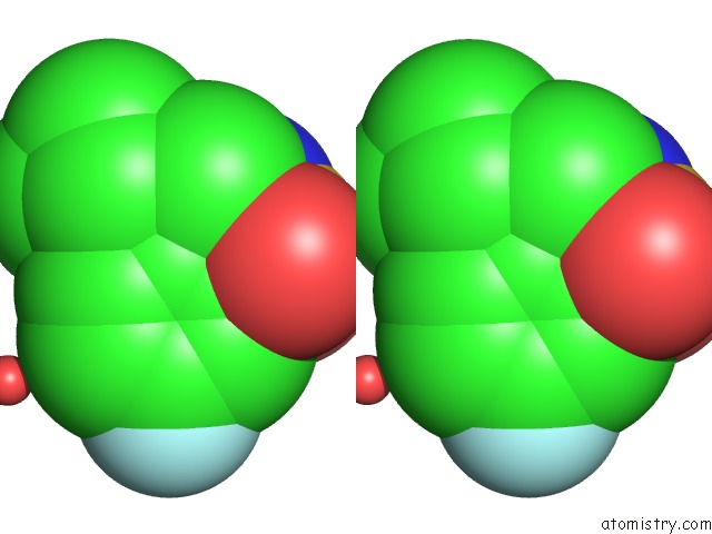
Stereo pair view
Mono view
Stereo pair view
A full contact list of Fluorine with other atoms in the F binding
site number 1 of Crystal Structure of the GLUA2 Ligand-Binding Domain (S1S2J-L483Y- N754S) in Complex with Glutamate and Bpam-344 at 1.87 A Resolution within 5.0Å range:
|
Fluorine binding site 2 out of 3 in 4n07
Go back to
Fluorine binding site 2 out
of 3 in the Crystal Structure of the GLUA2 Ligand-Binding Domain (S1S2J-L483Y- N754S) in Complex with Glutamate and Bpam-344 at 1.87 A Resolution
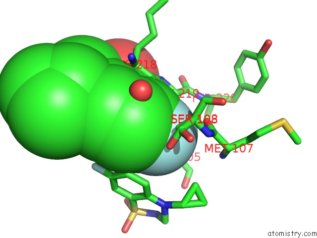
Mono view
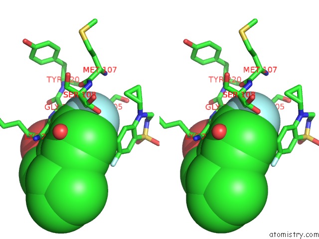
Stereo pair view
Mono view
Stereo pair view
A full contact list of Fluorine with other atoms in the F binding
site number 2 of Crystal Structure of the GLUA2 Ligand-Binding Domain (S1S2J-L483Y- N754S) in Complex with Glutamate and Bpam-344 at 1.87 A Resolution within 5.0Å range:
|
Fluorine binding site 3 out of 3 in 4n07
Go back to
Fluorine binding site 3 out
of 3 in the Crystal Structure of the GLUA2 Ligand-Binding Domain (S1S2J-L483Y- N754S) in Complex with Glutamate and Bpam-344 at 1.87 A Resolution
Mono view
Stereo pair view
Mono view
Stereo pair view
A full contact list of Fluorine with other atoms in the F binding
site number 3 of Crystal Structure of the GLUA2 Ligand-Binding Domain (S1S2J-L483Y- N754S) in Complex with Glutamate and Bpam-344 at 1.87 A Resolution within 5.0Å range:
|
Reference:
A.B.Nrholm,
P.Francotte,
L.Olsen,
C.Krintel,
K.Frydenvang,
E.Goffin,
S.Challal,
L.Danober,
I.Botez-Pop,
P.Lestage,
B.Pirotte,
J.S.Kastrup.
Synthesis, Pharmacological and Structural Characterization, and Thermodynamic Aspects of GLUA2-Positive Allosteric Modulators with A 3,4-Dihydro-2H-1,2,4-Benzothiadiazine 1,1-Dioxide Scaffold. J.Med.Chem. V. 56 8736 2013.
ISSN: ISSN 0022-2623
PubMed: 24131202
DOI: 10.1021/JM4012092
Page generated: Mon Jul 14 23:32:30 2025
ISSN: ISSN 0022-2623
PubMed: 24131202
DOI: 10.1021/JM4012092
Last articles
Fe in 2YXOFe in 2YRS
Fe in 2YXC
Fe in 2YNM
Fe in 2YVJ
Fe in 2YP1
Fe in 2YU2
Fe in 2YU1
Fe in 2YQB
Fe in 2YOO