Fluorine »
PDB 4o28-4olh »
4o5e »
Fluorine in PDB 4o5e: Structure of Human Dna Polymerase Complexed with N7MG in the Template Base Paired with Incoming Non-Hydrolyzable Ttp
Enzymatic activity of Structure of Human Dna Polymerase Complexed with N7MG in the Template Base Paired with Incoming Non-Hydrolyzable Ttp
All present enzymatic activity of Structure of Human Dna Polymerase Complexed with N7MG in the Template Base Paired with Incoming Non-Hydrolyzable Ttp:
2.7.7.7;
2.7.7.7;
Protein crystallography data
The structure of Structure of Human Dna Polymerase Complexed with N7MG in the Template Base Paired with Incoming Non-Hydrolyzable Ttp, PDB code: 4o5e
was solved by
M-.C.Koag,
S.Lee,
with X-Ray Crystallography technique. A brief refinement statistics is given in the table below:
| Resolution Low / High (Å) | 19.73 / 2.53 |
| Space group | P 1 21 1 |
| Cell size a, b, c (Å), α, β, γ (°) | 54.425, 78.920, 54.819, 90.00, 105.97, 90.00 |
| R / Rfree (%) | 20.5 / 27.3 |
Other elements in 4o5e:
The structure of Structure of Human Dna Polymerase Complexed with N7MG in the Template Base Paired with Incoming Non-Hydrolyzable Ttp also contains other interesting chemical elements:
| Magnesium | (Mg) | 1 atom |
| Sodium | (Na) | 2 atoms |
Fluorine Binding Sites:
The binding sites of Fluorine atom in the Structure of Human Dna Polymerase Complexed with N7MG in the Template Base Paired with Incoming Non-Hydrolyzable Ttp
(pdb code 4o5e). This binding sites where shown within
5.0 Angstroms radius around Fluorine atom.
In total only one binding site of Fluorine was determined in the Structure of Human Dna Polymerase Complexed with N7MG in the Template Base Paired with Incoming Non-Hydrolyzable Ttp, PDB code: 4o5e:
In total only one binding site of Fluorine was determined in the Structure of Human Dna Polymerase Complexed with N7MG in the Template Base Paired with Incoming Non-Hydrolyzable Ttp, PDB code: 4o5e:
Fluorine binding site 1 out of 1 in 4o5e
Go back to
Fluorine binding site 1 out
of 1 in the Structure of Human Dna Polymerase Complexed with N7MG in the Template Base Paired with Incoming Non-Hydrolyzable Ttp
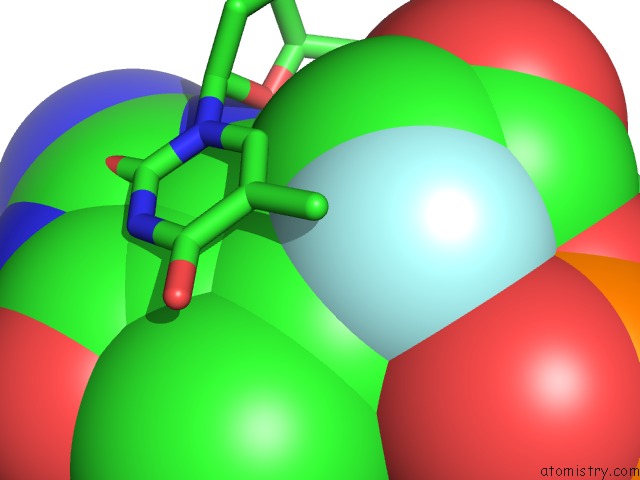
Mono view
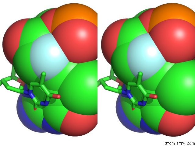
Stereo pair view
Mono view
Stereo pair view
A full contact list of Fluorine with other atoms in the F binding
site number 1 of Structure of Human Dna Polymerase Complexed with N7MG in the Template Base Paired with Incoming Non-Hydrolyzable Ttp within 5.0Å range:
|
Reference:
M.C.Koag,
Y.Kou,
H.Ouzon-Shubeita,
S.Lee.
Transition-State Destabilization Reveals How Human Dna Polymerase Beta Proceeds Across the Chemically Unstable Lesion N7-Methylguanine. Nucleic Acids Res. V. 42 8755 2014.
ISSN: ISSN 0305-1048
PubMed: 24966350
DOI: 10.1093/NAR/GKU554
Page generated: Mon Jul 14 23:48:32 2025
ISSN: ISSN 0305-1048
PubMed: 24966350
DOI: 10.1093/NAR/GKU554
Last articles
Fe in 2YXOFe in 2YRS
Fe in 2YXC
Fe in 2YNM
Fe in 2YVJ
Fe in 2YP1
Fe in 2YU2
Fe in 2YU1
Fe in 2YQB
Fe in 2YOO