Fluorine »
PDB 4o28-4olh »
4o91 »
Fluorine in PDB 4o91: Crystal Structure of Type II Inhibitor NG25 Bound to TAK1-TAB1
Enzymatic activity of Crystal Structure of Type II Inhibitor NG25 Bound to TAK1-TAB1
All present enzymatic activity of Crystal Structure of Type II Inhibitor NG25 Bound to TAK1-TAB1:
2.7.11.25;
2.7.11.25;
Protein crystallography data
The structure of Crystal Structure of Type II Inhibitor NG25 Bound to TAK1-TAB1, PDB code: 4o91
was solved by
D.Gurbani,
J.C.Hunter,
L.Tan,
K.D.Westover,
with X-Ray Crystallography technique. A brief refinement statistics is given in the table below:
| Resolution Low / High (Å) | 35.25 / 2.39 |
| Space group | I 2 2 2 |
| Cell size a, b, c (Å), α, β, γ (°) | 58.214, 132.905, 145.498, 90.00, 90.00, 90.00 |
| R / Rfree (%) | 17.7 / 20.7 |
Fluorine Binding Sites:
The binding sites of Fluorine atom in the Crystal Structure of Type II Inhibitor NG25 Bound to TAK1-TAB1
(pdb code 4o91). This binding sites where shown within
5.0 Angstroms radius around Fluorine atom.
In total 3 binding sites of Fluorine where determined in the Crystal Structure of Type II Inhibitor NG25 Bound to TAK1-TAB1, PDB code: 4o91:
Jump to Fluorine binding site number: 1; 2; 3;
In total 3 binding sites of Fluorine where determined in the Crystal Structure of Type II Inhibitor NG25 Bound to TAK1-TAB1, PDB code: 4o91:
Jump to Fluorine binding site number: 1; 2; 3;
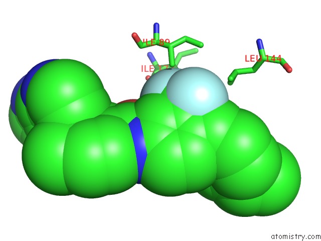
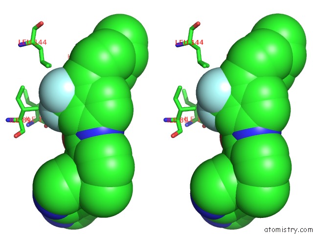
Fluorine binding site 1 out of 3 in 4o91
Go back to
Fluorine binding site 1 out
of 3 in the Crystal Structure of Type II Inhibitor NG25 Bound to TAK1-TAB1
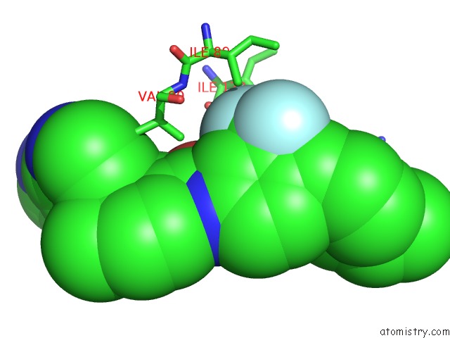
Mono view
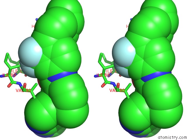
Stereo pair view
Mono view
Stereo pair view
A full contact list of Fluorine with other atoms in the F binding
site number 1 of Crystal Structure of Type II Inhibitor NG25 Bound to TAK1-TAB1 within 5.0Å range:
|
Fluorine binding site 2 out of 3 in 4o91
Go back to
Fluorine binding site 2 out
of 3 in the Crystal Structure of Type II Inhibitor NG25 Bound to TAK1-TAB1
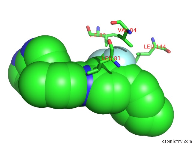
Mono view
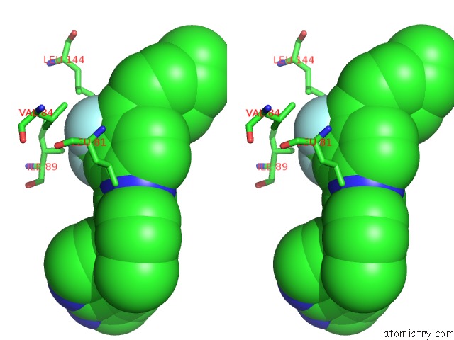
Stereo pair view
Mono view
Stereo pair view
A full contact list of Fluorine with other atoms in the F binding
site number 2 of Crystal Structure of Type II Inhibitor NG25 Bound to TAK1-TAB1 within 5.0Å range:
|
Fluorine binding site 3 out of 3 in 4o91
Go back to
Fluorine binding site 3 out
of 3 in the Crystal Structure of Type II Inhibitor NG25 Bound to TAK1-TAB1
Mono view
Stereo pair view
Mono view
Stereo pair view
A full contact list of Fluorine with other atoms in the F binding
site number 3 of Crystal Structure of Type II Inhibitor NG25 Bound to TAK1-TAB1 within 5.0Å range:
|
Reference:
L.Tan,
T.Nomanbhoy,
D.Gurbani,
M.Patricelli,
J.Hunter,
J.Geng,
L.Herhaus,
J.Zhang,
E.Pauls,
Y.Ham,
H.G.Choi,
T.Xie,
X.Deng,
S.J.Buhrlage,
T.Sim,
P.Cohen,
G.Sapkota,
K.D.Westover,
N.S.Gray.
Discovery of Type II Inhibitors of Tgf Beta-Activated Kinase 1 (TAK1) and Mitogen-Activated Protein Kinase Kinase Kinase Kinase 2 (MAP4K2). J.Med.Chem. 2014.
ISSN: ISSN 0022-2623
PubMed: 25075558
DOI: 10.1021/JM500480K
Page generated: Thu Aug 1 04:30:07 2024
ISSN: ISSN 0022-2623
PubMed: 25075558
DOI: 10.1021/JM500480K
Last articles
Zn in 9J0NZn in 9J0O
Zn in 9J0P
Zn in 9FJX
Zn in 9EKB
Zn in 9C0F
Zn in 9CAH
Zn in 9CH0
Zn in 9CH3
Zn in 9CH1