Fluorine »
PDB 4olm-4pa0 »
4p5z »
Fluorine in PDB 4p5z: Human EPHA3 Kinase Domain in Complex with Quinoxaline Derivatives
Enzymatic activity of Human EPHA3 Kinase Domain in Complex with Quinoxaline Derivatives
All present enzymatic activity of Human EPHA3 Kinase Domain in Complex with Quinoxaline Derivatives:
2.7.10.1;
2.7.10.1;
Protein crystallography data
The structure of Human EPHA3 Kinase Domain in Complex with Quinoxaline Derivatives, PDB code: 4p5z
was solved by
J.Dong,
A.Caflisch,
with X-Ray Crystallography technique. A brief refinement statistics is given in the table below:
| Resolution Low / High (Å) | 47.02 / 2.00 |
| Space group | P 1 21 1 |
| Cell size a, b, c (Å), α, β, γ (°) | 52.805, 38.235, 75.465, 90.00, 101.46, 90.00 |
| R / Rfree (%) | 17.8 / 21.9 |
Fluorine Binding Sites:
The binding sites of Fluorine atom in the Human EPHA3 Kinase Domain in Complex with Quinoxaline Derivatives
(pdb code 4p5z). This binding sites where shown within
5.0 Angstroms radius around Fluorine atom.
In total 3 binding sites of Fluorine where determined in the Human EPHA3 Kinase Domain in Complex with Quinoxaline Derivatives, PDB code: 4p5z:
Jump to Fluorine binding site number: 1; 2; 3;
In total 3 binding sites of Fluorine where determined in the Human EPHA3 Kinase Domain in Complex with Quinoxaline Derivatives, PDB code: 4p5z:
Jump to Fluorine binding site number: 1; 2; 3;
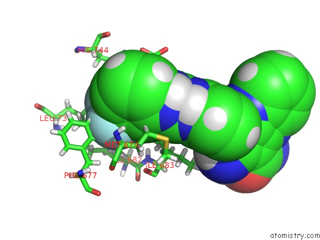
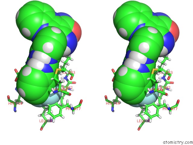
Fluorine binding site 1 out of 3 in 4p5z
Go back to
Fluorine binding site 1 out
of 3 in the Human EPHA3 Kinase Domain in Complex with Quinoxaline Derivatives
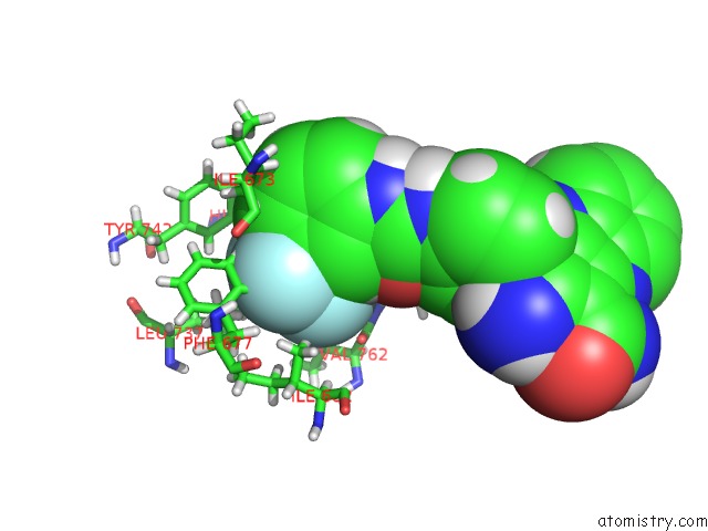
Mono view
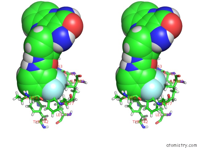
Stereo pair view
Mono view
Stereo pair view
A full contact list of Fluorine with other atoms in the F binding
site number 1 of Human EPHA3 Kinase Domain in Complex with Quinoxaline Derivatives within 5.0Å range:
|
Fluorine binding site 2 out of 3 in 4p5z
Go back to
Fluorine binding site 2 out
of 3 in the Human EPHA3 Kinase Domain in Complex with Quinoxaline Derivatives
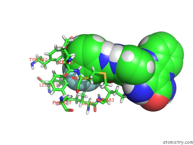
Mono view
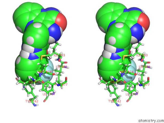
Stereo pair view
Mono view
Stereo pair view
A full contact list of Fluorine with other atoms in the F binding
site number 2 of Human EPHA3 Kinase Domain in Complex with Quinoxaline Derivatives within 5.0Å range:
|
Fluorine binding site 3 out of 3 in 4p5z
Go back to
Fluorine binding site 3 out
of 3 in the Human EPHA3 Kinase Domain in Complex with Quinoxaline Derivatives
Mono view
Stereo pair view
Mono view
Stereo pair view
A full contact list of Fluorine with other atoms in the F binding
site number 3 of Human EPHA3 Kinase Domain in Complex with Quinoxaline Derivatives within 5.0Å range:
|
Reference:
A.Unzue,
J.Dong,
K.Lafleur,
H.Zhao,
E.Frugier,
A.Caflisch,
C.Nevado.
Pyrrolo[3,2-B]Quinoxaline Derivatives As Types I1/2 and II Eph Tyrosine Kinase Inhibitors: Structure-Based Design, Synthesis, and in Vivo Validation. J.Med.Chem. V. 57 6834 2014.
ISSN: ISSN 0022-2623
PubMed: 25076195
DOI: 10.1021/JM5009242
Page generated: Thu Aug 1 04:46:07 2024
ISSN: ISSN 0022-2623
PubMed: 25076195
DOI: 10.1021/JM5009242
Last articles
Zn in 9J0NZn in 9J0O
Zn in 9J0P
Zn in 9FJX
Zn in 9EKB
Zn in 9C0F
Zn in 9CAH
Zn in 9CH0
Zn in 9CH3
Zn in 9CH1