Atomistry » Fluorine » PDB 4pb2-4q0d » 4pjt
Atomistry »
  Fluorine »
    PDB 4pb2-4q0d »
      4pjt »

Fluorine in PDB 4pjt: Structure of PARP1 Catalytic Domain Bound to Inhibitor Bmn 673

Enzymatic activity of Structure of PARP1 Catalytic Domain Bound to Inhibitor Bmn 673

All present enzymatic activity of Structure of PARP1 Catalytic Domain Bound to Inhibitor Bmn 673:
2.4.2.30;

Protein crystallography data

The structure of Structure of PARP1 Catalytic Domain Bound to Inhibitor Bmn 673, PDB code: 4pjt was solved by M.Aoyagi-Scharber, A.S.Gardberg, T.L.Arakaki, with X-Ray Crystallography technique. A brief refinement statistics is given in the table below:

Resolution Low / High (Å) 19.94 / 2.35
Space group P 21 21 21
Cell size a, b, c (Å), α, β, γ (°) 103.690, 108.150, 142.000, 90.00, 90.00, 90.00
R / Rfree (%) 18.8 / 22.8

Fluorine Binding Sites:

The binding sites of Fluorine atom in the Structure of PARP1 Catalytic Domain Bound to Inhibitor Bmn 673 (pdb code 4pjt). This binding sites where shown within 5.0 Angstroms radius around Fluorine atom.
In total 8 binding sites of Fluorine where determined in the Structure of PARP1 Catalytic Domain Bound to Inhibitor Bmn 673, PDB code: 4pjt:
Jump to Fluorine binding site number: 1; 2; 3; 4; 5; 6; 7; 8;

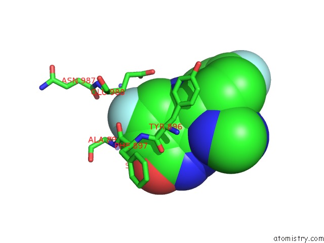
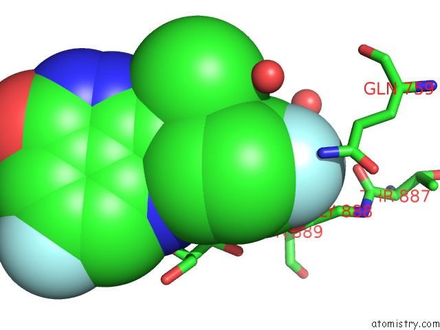
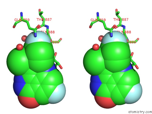
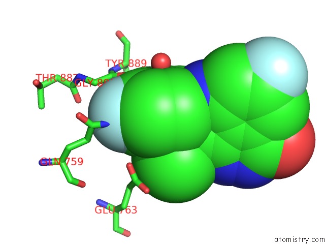
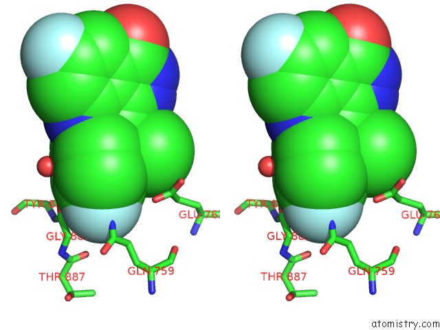
Fluorine binding site 1 out of 8 in 4pjt

Go back to Fluorine Binding Sites List in 4pjt
Fluorine binding site 1 out of 8 in the Structure of PARP1 Catalytic Domain Bound to Inhibitor Bmn 673


Mono view


Stereo pair view

A full contact list of Fluorine with other atoms in the F binding site number 1 of Structure of PARP1 Catalytic Domain Bound to Inhibitor Bmn 673 within 5.0Å range:
probe atom residue distance (Å) B Occ
A:F1105

b:24.4
occ:1.00
F2 A:2YQ1105 0.0 24.4 1.0
C14 A:2YQ1105 1.3 23.2 1.0
C15 A:2YQ1105 2.3 23.4 1.0
C13 A:2YQ1105 2.4 22.6 1.0
O A:GLY888 3.0 25.7 1.0
C A:GLY888 3.1 25.4 1.0
CA A:GLY888 3.3 28.2 1.0
O A:HOH1280 3.3 46.1 1.0
C16 A:2YQ1105 3.6 21.6 1.0
C12 A:2YQ1105 3.6 22.4 1.0
NE2 A:GLN759 3.7 61.2 1.0
N A:TYR889 3.9 25.5 1.0
OE2 A:GLU763 3.9 57.9 1.0
CG A:GLN759 4.0 52.5 1.0
CD A:GLN759 4.0 63.0 1.0
C11 A:2YQ1105 4.1 21.0 1.0
O A:THR887 4.3 36.4 1.0
N A:GLY888 4.5 32.8 1.0
CA A:TYR889 4.6 27.6 1.0
O A:HOH1232 4.7 26.9 1.0
CD1 A:TYR889 4.7 33.9 1.0
C A:THR887 4.9 37.9 1.0
CG A:TYR889 4.9 29.8 1.0
CE1 A:TYR889 4.9 35.6 1.0
OE1 A:GLN759 4.9 69.0 1.0
O A:HOH1274 4.9 33.2 1.0

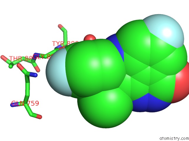
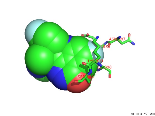
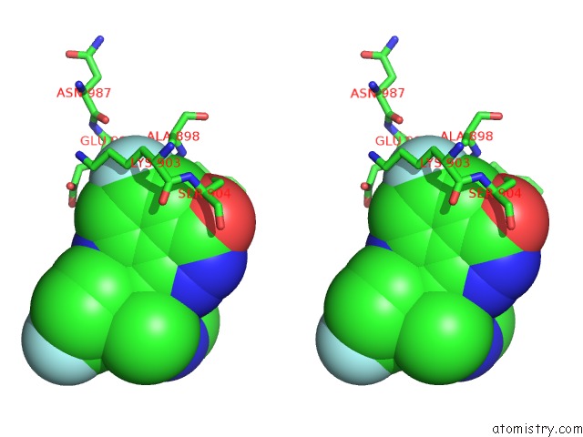
Fluorine binding site 2 out of 8 in 4pjt

Go back to Fluorine Binding Sites List in 4pjt
Fluorine binding site 2 out of 8 in the Structure of PARP1 Catalytic Domain Bound to Inhibitor Bmn 673


Mono view


Stereo pair view

A full contact list of Fluorine with other atoms in the F binding site number 2 of Structure of PARP1 Catalytic Domain Bound to Inhibitor Bmn 673 within 5.0Å range:
probe atom residue distance (Å) B Occ
A:F1105

b:20.9
occ:1.00
F1 A:2YQ1105 0.0 20.9 1.0
C9 A:2YQ1105 1.3 19.6 1.0
C8 A:2YQ1105 2.3 19.3 1.0
C10 A:2YQ1105 2.4 19.3 1.0
CB A:GLU988 3.4 20.4 1.0
O A:PHE897 3.4 18.2 1.0
CE A:LYS903 3.5 22.4 1.0
C7 A:2YQ1105 3.6 19.0 1.0
C1 A:2YQ1105 3.6 19.6 1.0
CB A:ALA898 3.6 17.6 1.0
C A:PHE897 3.7 17.5 1.0
CD A:LYS903 3.8 22.3 1.0
CG A:LYS903 3.8 22.3 1.0
N A:ALA898 3.9 16.6 1.0
CA A:ALA898 3.9 17.0 1.0
C2 A:2YQ1105 4.1 19.4 1.0
NZ A:LYS903 4.1 24.2 1.0
OE2 A:GLU988 4.1 21.2 1.0
CD A:GLU988 4.3 20.3 1.0
CA A:GLU988 4.3 20.2 1.0
CG A:GLU988 4.4 20.6 1.0
O A:ASN987 4.5 19.2 1.0
N A:PHE897 4.5 18.1 1.0
CA A:PHE897 4.5 17.6 1.0
N3 A:2YQ1105 4.7 18.5 1.0
O A:HOH1236 4.7 32.1 1.0
OE1 A:GLU988 4.8 19.6 1.0
C4 A:2YQ1105 4.8 19.3 1.0
CB A:LYS903 4.8 21.4 1.0
OG A:SER904 4.9 21.2 1.0
CB A:TYR896 5.0 20.1 1.0

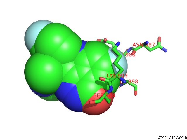
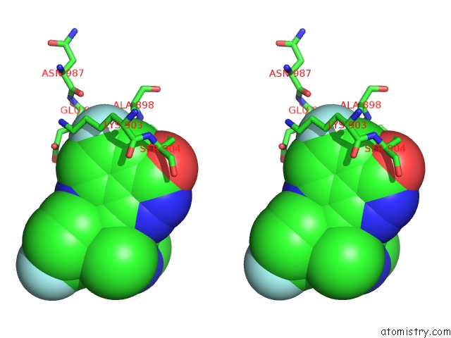
Fluorine binding site 3 out of 8 in 4pjt

Go back to Fluorine Binding Sites List in 4pjt
Fluorine binding site 3 out of 8 in the Structure of PARP1 Catalytic Domain Bound to Inhibitor Bmn 673


Mono view


Stereo pair view

A full contact list of Fluorine with other atoms in the F binding site number 3 of Structure of PARP1 Catalytic Domain Bound to Inhibitor Bmn 673 within 5.0Å range:
probe atom residue distance (Å) B Occ
B:F1105

b:45.4
occ:1.00
F2 B:2YQ1105 0.0 45.4 1.0
C14 B:2YQ1105 1.3 40.7 1.0
C15 B:2YQ1105 2.3 35.0 1.0
C13 B:2YQ1105 2.4 37.9 1.0
O B:GLY888 2.9 47.8 1.0
C B:GLY888 3.1 49.0 1.0
CA B:GLY888 3.5 49.8 1.0
C16 B:2YQ1105 3.6 34.5 1.0
C12 B:2YQ1105 3.6 35.7 1.0
N B:TYR889 3.9 48.4 1.0
C11 B:2YQ1105 4.1 31.7 1.0
O B:HOH1210 4.3 40.2 1.0
CA B:TYR889 4.5 45.7 1.0
O B:THR887 4.5 51.6 1.0
N B:GLY888 4.7 51.4 1.0
NE2 B:GLN759 4.8 61.6 1.0
CG B:TYR889 5.0 45.0 1.0

Fluorine binding site 4 out of 8 in 4pjt

Go back to Fluorine Binding Sites List in 4pjt
Fluorine binding site 4 out of 8 in the Structure of PARP1 Catalytic Domain Bound to Inhibitor Bmn 673


Mono view


Stereo pair view

A full contact list of Fluorine with other atoms in the F binding site number 4 of Structure of PARP1 Catalytic Domain Bound to Inhibitor Bmn 673 within 5.0Å range:
probe atom residue distance (Å) B Occ
B:F1105

b:27.6
occ:1.00
F1 B:2YQ1105 0.0 27.6 1.0
C9 B:2YQ1105 1.3 24.5 1.0
C10 B:2YQ1105 2.3 23.6 1.0
C8 B:2YQ1105 2.3 23.8 1.0
O B:PHE897 3.3 21.7 1.0
CB B:GLU988 3.3 31.4 1.0
CE B:LYS903 3.4 32.6 1.0
CB B:ALA898 3.5 26.0 1.0
C B:PHE897 3.6 23.0 1.0
C7 B:2YQ1105 3.6 24.5 1.0
C1 B:2YQ1105 3.6 23.7 1.0
CD B:LYS903 3.7 31.2 1.0
N B:ALA898 3.7 24.4 1.0
CG B:LYS903 3.8 28.9 1.0
CA B:ALA898 3.8 24.1 1.0
NZ B:LYS903 4.0 33.1 1.0
C2 B:2YQ1105 4.1 25.2 1.0
CA B:GLU988 4.1 30.6 1.0
OE2 B:GLU988 4.1 39.5 1.0
O B:ASN987 4.2 31.4 1.0
CD B:GLU988 4.3 36.0 1.0
CG B:GLU988 4.4 33.6 1.0
CA B:PHE897 4.5 21.8 1.0
N B:PHE897 4.5 22.8 1.0
N3 B:2YQ1105 4.7 26.1 1.0
C4 B:2YQ1105 4.8 24.5 1.0
CB B:LYS903 4.8 29.9 1.0
OG B:SER904 4.9 29.1 1.0
OE1 B:GLU988 4.9 39.4 1.0

Fluorine binding site 5 out of 8 in 4pjt

Go back to Fluorine Binding Sites List in 4pjt
Fluorine binding site 5 out of 8 in the Structure of PARP1 Catalytic Domain Bound to Inhibitor Bmn 673


Mono view


Stereo pair view

A full contact list of Fluorine with other atoms in the F binding site number 5 of Structure of PARP1 Catalytic Domain Bound to Inhibitor Bmn 673 within 5.0Å range:
probe atom residue distance (Å) B Occ
C:F1104

b:28.6
occ:1.00
F2 C:2YQ1104 0.0 28.6 1.0
C14 C:2YQ1104 1.3 24.9 1.0
C15 C:2YQ1104 2.3 22.7 1.0
C13 C:2YQ1104 2.3 23.7 1.0
O C:GLY888 3.0 29.0 1.0
C C:GLY888 3.1 27.3 1.0
NE2 C:GLN759 3.2 34.9 1.0
CA C:GLY888 3.4 27.4 1.0
C16 C:2YQ1104 3.6 21.9 1.0
C12 C:2YQ1104 3.6 22.2 1.0
CD C:GLN759 3.8 37.7 1.0
N C:TYR889 3.8 25.2 1.0
O C:HOH1241 4.0 33.0 1.0
C11 C:2YQ1104 4.1 20.3 1.0
CG C:GLN759 4.1 34.2 1.0
O C:THR887 4.1 28.8 1.0
O C:HOH1298 4.5 51.0 1.0
CA C:TYR889 4.5 26.5 1.0
N C:GLY888 4.6 28.8 1.0
O3 C:GOL1105 4.6 31.6 1.0
OE1 C:GLN759 4.7 40.5 1.0
C C:THR887 4.8 30.1 1.0
C3 C:GOL1105 4.9 35.1 1.0
CD1 C:TYR889 5.0 23.5 1.0
CG C:TYR889 5.0 24.5 1.0

Fluorine binding site 6 out of 8 in 4pjt

Go back to Fluorine Binding Sites List in 4pjt
Fluorine binding site 6 out of 8 in the Structure of PARP1 Catalytic Domain Bound to Inhibitor Bmn 673


Mono view


Stereo pair view

A full contact list of Fluorine with other atoms in the F binding site number 6 of Structure of PARP1 Catalytic Domain Bound to Inhibitor Bmn 673 within 5.0Å range:
probe atom residue distance (Å) B Occ
C:F1104

b:20.7
occ:1.00
F1 C:2YQ1104 0.0 20.7 1.0
C9 C:2YQ1104 1.4 19.8 1.0
C10 C:2YQ1104 2.4 18.9 1.0
C8 C:2YQ1104 2.4 18.5 1.0
O C:PHE897 3.3 18.2 1.0
CE C:LYS903 3.4 22.5 1.0
CB C:ALA898 3.5 21.7 1.0
CB C:GLU988 3.5 22.9 1.0
C C:PHE897 3.5 18.1 1.0
CD C:LYS903 3.6 22.5 1.0
C7 C:2YQ1104 3.6 18.9 1.0
C1 C:2YQ1104 3.6 19.0 1.0
N C:ALA898 3.7 19.9 1.0
CG C:LYS903 3.7 22.4 1.0
CA C:ALA898 3.7 20.5 1.0
NZ C:LYS903 4.1 23.1 1.0
C2 C:2YQ1104 4.1 18.9 1.0
OE2 C:GLU988 4.3 26.4 1.0
CA C:GLU988 4.3 21.0 1.0
O C:ASN987 4.3 19.9 1.0
CA C:PHE897 4.4 17.3 1.0
N C:PHE897 4.5 17.2 1.0
CD C:GLU988 4.5 25.7 1.0
CG C:GLU988 4.6 23.8 1.0
CB C:LYS903 4.7 23.0 1.0
OG C:SER904 4.8 19.9 1.0
N3 C:2YQ1104 4.8 17.8 1.0
C4 C:2YQ1104 4.8 19.5 1.0
O C:HOH1215 4.9 35.3 1.0
O2 C:GOL1105 5.0 28.1 1.0

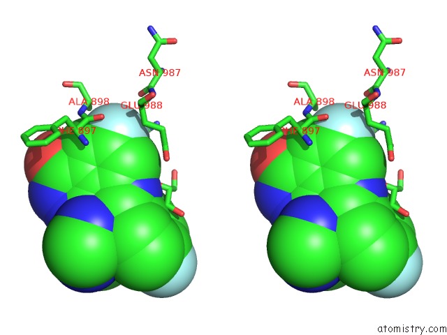
Fluorine binding site 7 out of 8 in 4pjt

Go back to Fluorine Binding Sites List in 4pjt
Fluorine binding site 7 out of 8 in the Structure of PARP1 Catalytic Domain Bound to Inhibitor Bmn 673


Mono view


Stereo pair view

A full contact list of Fluorine with other atoms in the F binding site number 7 of Structure of PARP1 Catalytic Domain Bound to Inhibitor Bmn 673 within 5.0Å range:
probe atom residue distance (Å) B Occ
D:F1104

b:31.1
occ:1.00
F2 D:2YQ1104 0.0 31.1 1.0
C14 D:2YQ1104 1.3 30.4 1.0
C15 D:2YQ1104 2.3 30.1 1.0
C13 D:2YQ1104 2.3 30.7 1.0
NE2 D:GLN759 2.7 62.3 1.0
O D:GLY888 2.9 36.2 1.0
C D:GLY888 3.2 34.3 1.0
CD D:GLN759 3.3 66.2 1.0
CA D:GLY888 3.4 35.9 1.0
C12 D:2YQ1104 3.6 31.1 1.0
C16 D:2YQ1104 3.6 29.2 1.0
CG D:GLN759 3.6 59.1 1.0
OE1 D:GLU763 3.9 62.8 1.0
N D:TYR889 4.0 34.7 1.0
C11 D:2YQ1104 4.1 28.5 1.0
OE1 D:GLN759 4.3 62.3 1.0
O D:THR887 4.4 42.2 1.0
O D:HOH1230 4.6 39.5 1.0
N D:GLY888 4.6 37.2 1.0
CA D:TYR889 4.7 36.3 1.0
C D:THR887 5.0 40.8 1.0

Fluorine binding site 8 out of 8 in 4pjt

Go back to Fluorine Binding Sites List in 4pjt
Fluorine binding site 8 out of 8 in the Structure of PARP1 Catalytic Domain Bound to Inhibitor Bmn 673


Mono view


Stereo pair view

A full contact list of Fluorine with other atoms in the F binding site number 8 of Structure of PARP1 Catalytic Domain Bound to Inhibitor Bmn 673 within 5.0Å range:
probe atom residue distance (Å) B Occ
D:F1104

b:28.3
occ:1.00
F1 D:2YQ1104 0.0 28.3 1.0
C9 D:2YQ1104 1.4 25.7 1.0
C8 D:2YQ1104 2.3 26.6 1.0
C10 D:2YQ1104 2.4 24.4 1.0
O D:PHE897 3.4 24.1 1.0
CE D:LYS903 3.4 31.1 1.0
CB D:GLU988 3.4 30.6 1.0
CB D:ALA898 3.5 29.8 1.0
C D:PHE897 3.6 26.8 1.0
C7 D:2YQ1104 3.6 27.3 1.0
CD D:LYS903 3.6 31.1 1.0
CG D:LYS903 3.6 31.5 1.0
C1 D:2YQ1104 3.7 24.8 1.0
N D:ALA898 3.8 27.8 1.0
CA D:ALA898 3.8 28.9 1.0
NZ D:LYS903 4.0 34.7 1.0
C2 D:2YQ1104 4.2 25.8 1.0
OE2 D:GLU988 4.2 35.0 1.0
CA D:GLU988 4.3 30.3 1.0
O D:ASN987 4.4 27.8 1.0
CD D:GLU988 4.4 33.0 1.0
CA D:PHE897 4.5 25.6 1.0
CG D:GLU988 4.5 31.6 1.0
N D:PHE897 4.5 26.4 1.0
CB D:LYS903 4.7 31.7 1.0
N3 D:2YQ1104 4.7 27.6 1.0
OG D:SER904 4.8 31.1 1.0
C4 D:2YQ1104 4.9 24.1 1.0
OE1 D:GLU988 5.0 33.3 1.0

Reference:

M.Aoyagi-Scharber, A.S.Gardberg, B.K.Yip, B.Wang, Y.Shen, P.A.Fitzpatrick. Structural Basis For the Inhibition of Poly(Adp-Ribose) Polymerases 1 and 2 By Bmn 673, A Potent Inhibitor Derived From Dihydropyridophthalazinone. Acta Crystallogr.,Sect.F V. 70 1143 2014.
ISSN: ESSN 2053-230X
PubMed: 25195882
DOI: 10.1107/S2053230X14015088
Page generated: Thu Aug 1 04:52:48 2024

Last articles

Zn in 9J0N
Zn in 9J0O
Zn in 9J0P
Zn in 9FJX
Zn in 9EKB
Zn in 9C0F
Zn in 9CAH
Zn in 9CH0
Zn in 9CH3
Zn in 9CH1
© Copyright 2008-2020 by atomistry.com
Home   |    Site Map   |    Copyright   |    Contact us   |    Privacy