Fluorine »
PDB 4pb2-4q0d »
4pvu »
Fluorine in PDB 4pvu: Crystal Structure of the Complex Between Ppargamma-Lbd and the R Enantiomer of Mbx-102 (Metaglidasen)
Protein crystallography data
The structure of Crystal Structure of the Complex Between Ppargamma-Lbd and the R Enantiomer of Mbx-102 (Metaglidasen), PDB code: 4pvu
was solved by
G.Pochetti,
R.Montanari,
D.Capelli,
F.Loiodice,
A.Laghezza,
L.Piemontese,
A.Lavecchia,
with X-Ray Crystallography technique. A brief refinement statistics is given in the table below:
| Resolution Low / High (Å) | 57.91 / 2.60 |
| Space group | C 1 2 1 |
| Cell size a, b, c (Å), α, β, γ (°) | 93.370, 60.210, 119.060, 90.00, 103.39, 90.00 |
| R / Rfree (%) | 20 / 25.1 |
Other elements in 4pvu:
The structure of Crystal Structure of the Complex Between Ppargamma-Lbd and the R Enantiomer of Mbx-102 (Metaglidasen) also contains other interesting chemical elements:
| Chlorine | (Cl) | 1 atom |
Fluorine Binding Sites:
The binding sites of Fluorine atom in the Crystal Structure of the Complex Between Ppargamma-Lbd and the R Enantiomer of Mbx-102 (Metaglidasen)
(pdb code 4pvu). This binding sites where shown within
5.0 Angstroms radius around Fluorine atom.
In total 3 binding sites of Fluorine where determined in the Crystal Structure of the Complex Between Ppargamma-Lbd and the R Enantiomer of Mbx-102 (Metaglidasen), PDB code: 4pvu:
Jump to Fluorine binding site number: 1; 2; 3;
In total 3 binding sites of Fluorine where determined in the Crystal Structure of the Complex Between Ppargamma-Lbd and the R Enantiomer of Mbx-102 (Metaglidasen), PDB code: 4pvu:
Jump to Fluorine binding site number: 1; 2; 3;
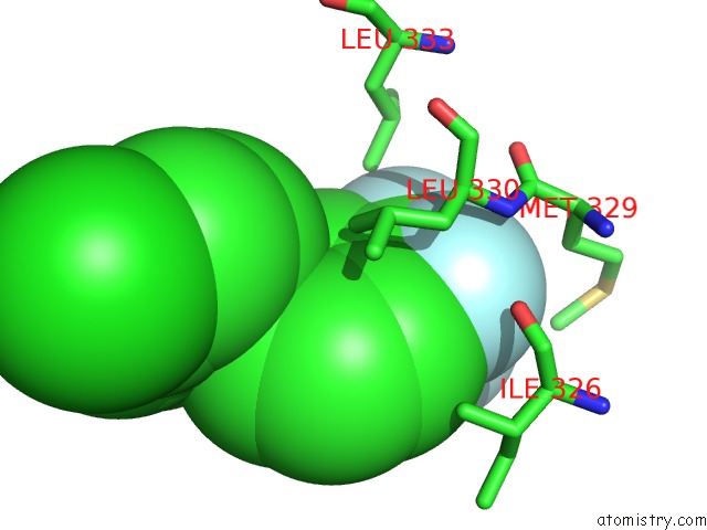
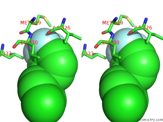
Fluorine binding site 1 out of 3 in 4pvu
Go back to
Fluorine binding site 1 out
of 3 in the Crystal Structure of the Complex Between Ppargamma-Lbd and the R Enantiomer of Mbx-102 (Metaglidasen)
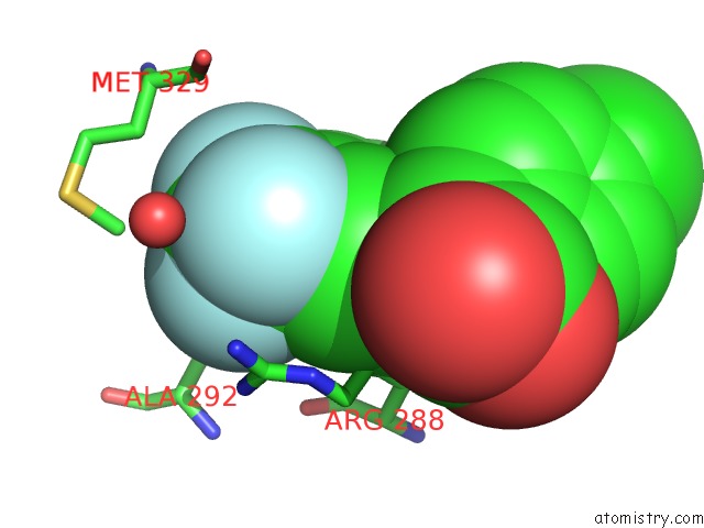
Mono view
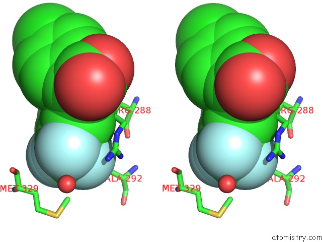
Stereo pair view
Mono view
Stereo pair view
A full contact list of Fluorine with other atoms in the F binding
site number 1 of Crystal Structure of the Complex Between Ppargamma-Lbd and the R Enantiomer of Mbx-102 (Metaglidasen) within 5.0Å range:
|
Fluorine binding site 2 out of 3 in 4pvu
Go back to
Fluorine binding site 2 out
of 3 in the Crystal Structure of the Complex Between Ppargamma-Lbd and the R Enantiomer of Mbx-102 (Metaglidasen)
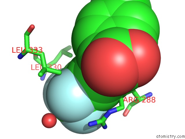
Mono view
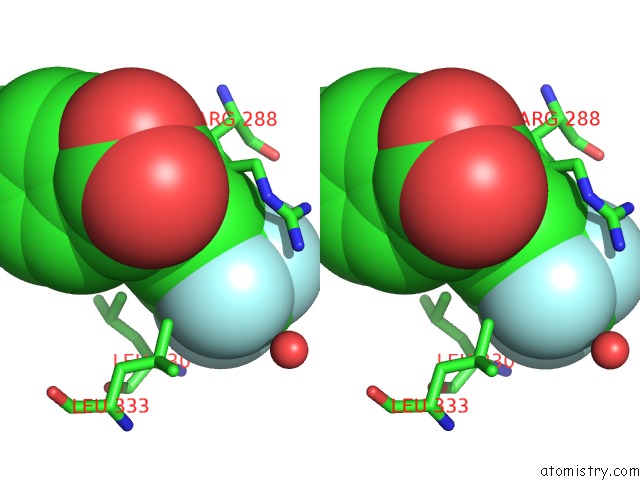
Stereo pair view
Mono view
Stereo pair view
A full contact list of Fluorine with other atoms in the F binding
site number 2 of Crystal Structure of the Complex Between Ppargamma-Lbd and the R Enantiomer of Mbx-102 (Metaglidasen) within 5.0Å range:
|
Fluorine binding site 3 out of 3 in 4pvu
Go back to
Fluorine binding site 3 out
of 3 in the Crystal Structure of the Complex Between Ppargamma-Lbd and the R Enantiomer of Mbx-102 (Metaglidasen)
Mono view
Stereo pair view
Mono view
Stereo pair view
A full contact list of Fluorine with other atoms in the F binding
site number 3 of Crystal Structure of the Complex Between Ppargamma-Lbd and the R Enantiomer of Mbx-102 (Metaglidasen) within 5.0Å range:
|
Reference:
A.Laghezza,
R.Montanari,
A.Lavecchia,
L.Piemontese,
G.Pochetti,
V.Iacobazzi,
V.Infantino,
D.Capelli,
M.De Bellis,
A.Liantonio,
S.Pierno,
P.Tortorella,
D.Conte Camerino,
F.Loiodice.
On the Metabolically Active Form of Metaglidasen: Improved Synthesis and Investigation of Its Peculiar Activity on Peroxisome Proliferator-Activated Receptors and Skeletal Muscles. Chemmedchem 2015.
ISSN: ESSN 1860-7187
PubMed: 25641779
DOI: 10.1002/CMDC.201402462
Page generated: Thu Aug 1 04:58:26 2024
ISSN: ESSN 1860-7187
PubMed: 25641779
DOI: 10.1002/CMDC.201402462
Last articles
Zn in 9J0NZn in 9J0O
Zn in 9J0P
Zn in 9FJX
Zn in 9EKB
Zn in 9C0F
Zn in 9CAH
Zn in 9CH0
Zn in 9CH3
Zn in 9CH1