Fluorine »
PDB 4q0e-4qrc »
4qjm »
Fluorine in PDB 4qjm: Crystal Structure of Human Carbonic Anhydrase Isozyme II with Inhibitor
Enzymatic activity of Crystal Structure of Human Carbonic Anhydrase Isozyme II with Inhibitor
All present enzymatic activity of Crystal Structure of Human Carbonic Anhydrase Isozyme II with Inhibitor:
4.2.1.1;
4.2.1.1;
Protein crystallography data
The structure of Crystal Structure of Human Carbonic Anhydrase Isozyme II with Inhibitor, PDB code: 4qjm
was solved by
A.Smirnov,
E.Manakova,
S.Grazulis,
with X-Ray Crystallography technique. A brief refinement statistics is given in the table below:
| Resolution Low / High (Å) | 20.55 / 1.75 |
| Space group | P 1 21 1 |
| Cell size a, b, c (Å), α, β, γ (°) | 42.187, 41.090, 71.981, 90.00, 104.12, 90.00 |
| R / Rfree (%) | 16.1 / 21.2 |
Other elements in 4qjm:
The structure of Crystal Structure of Human Carbonic Anhydrase Isozyme II with Inhibitor also contains other interesting chemical elements:
| Zinc | (Zn) | 1 atom |
Fluorine Binding Sites:
The binding sites of Fluorine atom in the Crystal Structure of Human Carbonic Anhydrase Isozyme II with Inhibitor
(pdb code 4qjm). This binding sites where shown within
5.0 Angstroms radius around Fluorine atom.
In total 3 binding sites of Fluorine where determined in the Crystal Structure of Human Carbonic Anhydrase Isozyme II with Inhibitor, PDB code: 4qjm:
Jump to Fluorine binding site number: 1; 2; 3;
In total 3 binding sites of Fluorine where determined in the Crystal Structure of Human Carbonic Anhydrase Isozyme II with Inhibitor, PDB code: 4qjm:
Jump to Fluorine binding site number: 1; 2; 3;
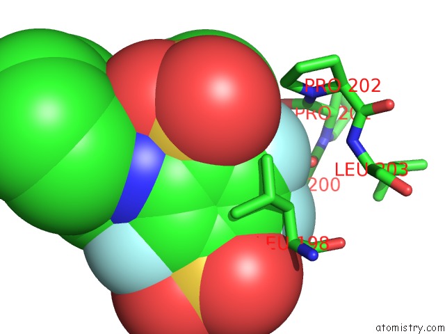
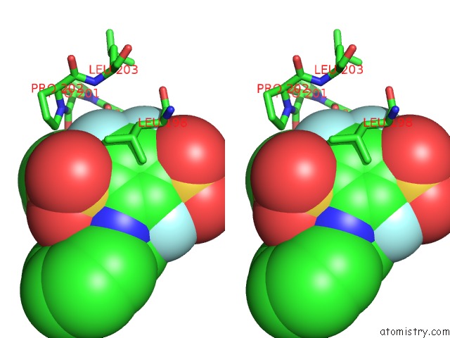
Fluorine binding site 1 out of 3 in 4qjm
Go back to
Fluorine binding site 1 out
of 3 in the Crystal Structure of Human Carbonic Anhydrase Isozyme II with Inhibitor
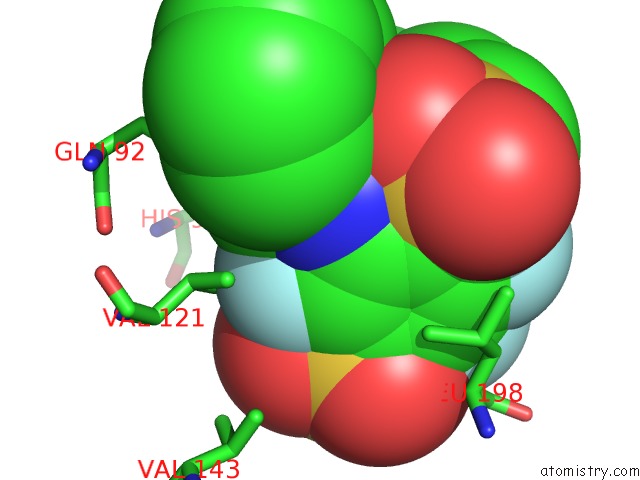
Mono view
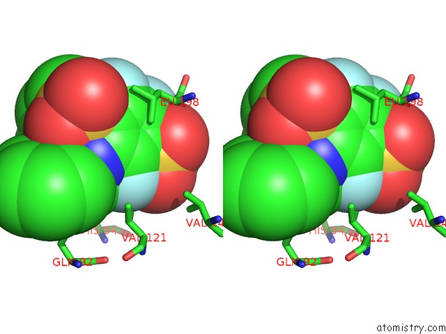
Stereo pair view
Mono view
Stereo pair view
A full contact list of Fluorine with other atoms in the F binding
site number 1 of Crystal Structure of Human Carbonic Anhydrase Isozyme II with Inhibitor within 5.0Å range:
|
Fluorine binding site 2 out of 3 in 4qjm
Go back to
Fluorine binding site 2 out
of 3 in the Crystal Structure of Human Carbonic Anhydrase Isozyme II with Inhibitor
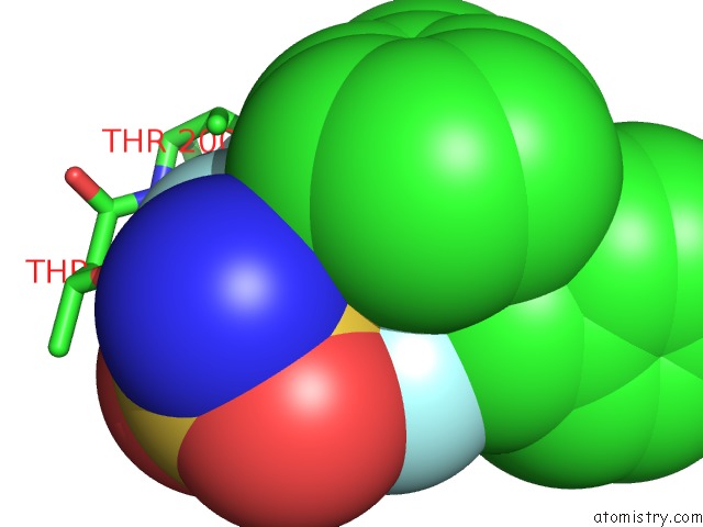
Mono view
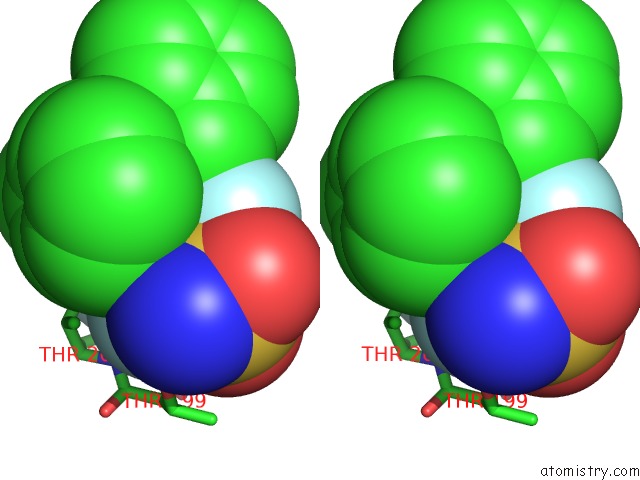
Stereo pair view
Mono view
Stereo pair view
A full contact list of Fluorine with other atoms in the F binding
site number 2 of Crystal Structure of Human Carbonic Anhydrase Isozyme II with Inhibitor within 5.0Å range:
|
Fluorine binding site 3 out of 3 in 4qjm
Go back to
Fluorine binding site 3 out
of 3 in the Crystal Structure of Human Carbonic Anhydrase Isozyme II with Inhibitor
Mono view
Stereo pair view
Mono view
Stereo pair view
A full contact list of Fluorine with other atoms in the F binding
site number 3 of Crystal Structure of Human Carbonic Anhydrase Isozyme II with Inhibitor within 5.0Å range:
|
Reference:
V.Dudutiene,
A.Zubriene,
A.Smirnov,
D.D.Timm,
J.Smirnoviene,
J.Kazokaite,
V.Michailoviene,
A.Zaksauskas,
E.Manakova,
S.Grazulis,
D.Matulis.
Functionalization of Fluorinated Benzenesulfonamides and Their Inhibitory Properties Toward Carbonic Anhydrases Chemmedchem V. 10 662 2015.
ISSN: ISSN 1860-7179
PubMed: 25758852
DOI: 10.1002/CMDC.201402490
Page generated: Thu Aug 1 05:16:04 2024
ISSN: ISSN 1860-7179
PubMed: 25758852
DOI: 10.1002/CMDC.201402490
Last articles
Zn in 9J0NZn in 9J0O
Zn in 9J0P
Zn in 9FJX
Zn in 9EKB
Zn in 9C0F
Zn in 9CAH
Zn in 9CH0
Zn in 9CH3
Zn in 9CH1