Fluorine »
PDB 4qte-4rv6 »
4rv3 »
Fluorine in PDB 4rv3: Crystal Structure of A Pentafluoro-Phe Incorporated Phosphatidylinositol-Specific Phospholipase C (H258X)From Staphylococcus Aureus
Enzymatic activity of Crystal Structure of A Pentafluoro-Phe Incorporated Phosphatidylinositol-Specific Phospholipase C (H258X)From Staphylococcus Aureus
All present enzymatic activity of Crystal Structure of A Pentafluoro-Phe Incorporated Phosphatidylinositol-Specific Phospholipase C (H258X)From Staphylococcus Aureus:
4.6.1.13;
4.6.1.13;
Protein crystallography data
The structure of Crystal Structure of A Pentafluoro-Phe Incorporated Phosphatidylinositol-Specific Phospholipase C (H258X)From Staphylococcus Aureus, PDB code: 4rv3
was solved by
T.He,
A.Gershenson,
S.J.Eyles,
J.Gao,
M.F.Roberts,
with X-Ray Crystallography technique. A brief refinement statistics is given in the table below:
| Resolution Low / High (Å) | 49.87 / 2.00 |
| Space group | P 43 21 2 |
| Cell size a, b, c (Å), α, β, γ (°) | 59.850, 59.850, 180.410, 90.00, 90.00, 90.00 |
| R / Rfree (%) | 18 / 23.8 |
Fluorine Binding Sites:
The binding sites of Fluorine atom in the Crystal Structure of A Pentafluoro-Phe Incorporated Phosphatidylinositol-Specific Phospholipase C (H258X)From Staphylococcus Aureus
(pdb code 4rv3). This binding sites where shown within
5.0 Angstroms radius around Fluorine atom.
In total 5 binding sites of Fluorine where determined in the Crystal Structure of A Pentafluoro-Phe Incorporated Phosphatidylinositol-Specific Phospholipase C (H258X)From Staphylococcus Aureus, PDB code: 4rv3:
Jump to Fluorine binding site number: 1; 2; 3; 4; 5;
In total 5 binding sites of Fluorine where determined in the Crystal Structure of A Pentafluoro-Phe Incorporated Phosphatidylinositol-Specific Phospholipase C (H258X)From Staphylococcus Aureus, PDB code: 4rv3:
Jump to Fluorine binding site number: 1; 2; 3; 4; 5;
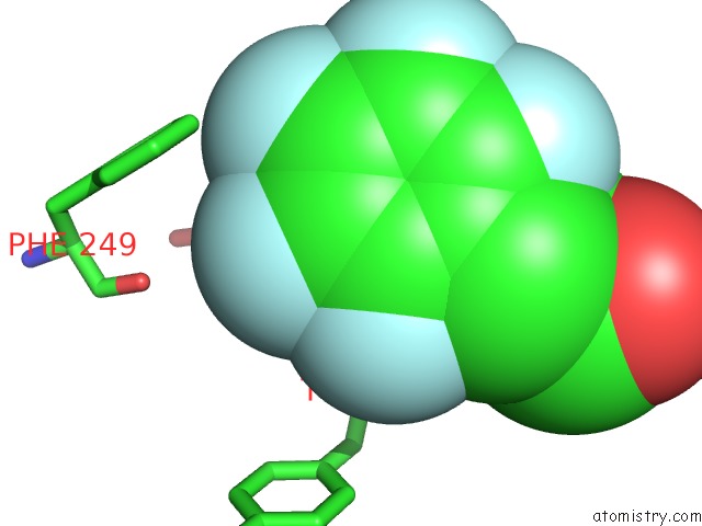
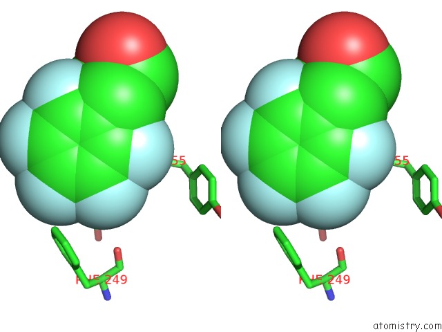
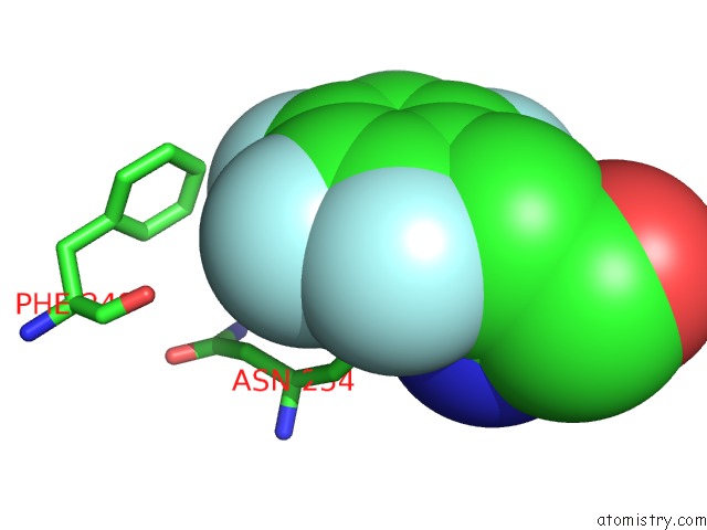
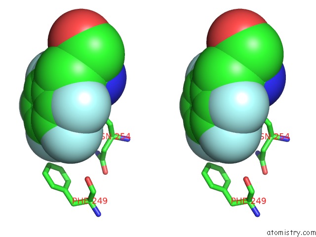
Fluorine binding site 1 out of 5 in 4rv3
Go back to
Fluorine binding site 1 out
of 5 in the Crystal Structure of A Pentafluoro-Phe Incorporated Phosphatidylinositol-Specific Phospholipase C (H258X)From Staphylococcus Aureus
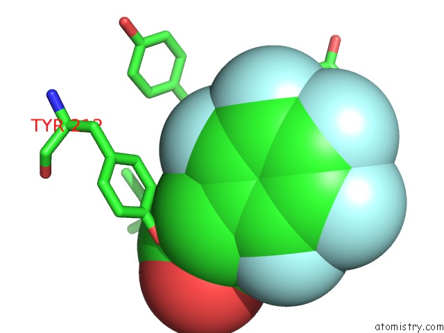
Mono view
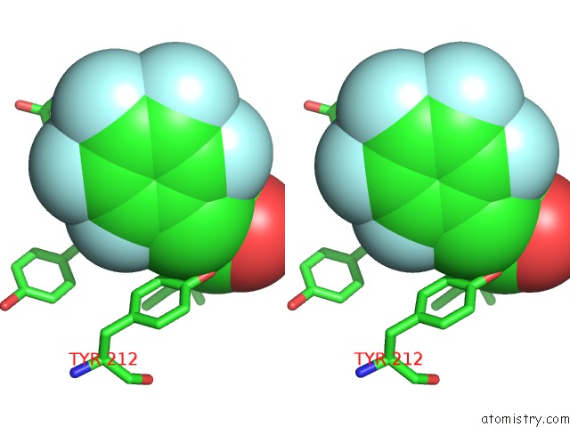
Stereo pair view
Mono view
Stereo pair view
A full contact list of Fluorine with other atoms in the F binding
site number 1 of Crystal Structure of A Pentafluoro-Phe Incorporated Phosphatidylinositol-Specific Phospholipase C (H258X)From Staphylococcus Aureus within 5.0Å range:
|
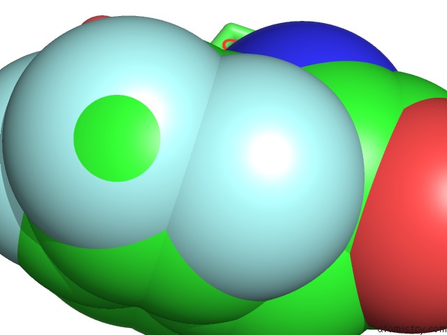
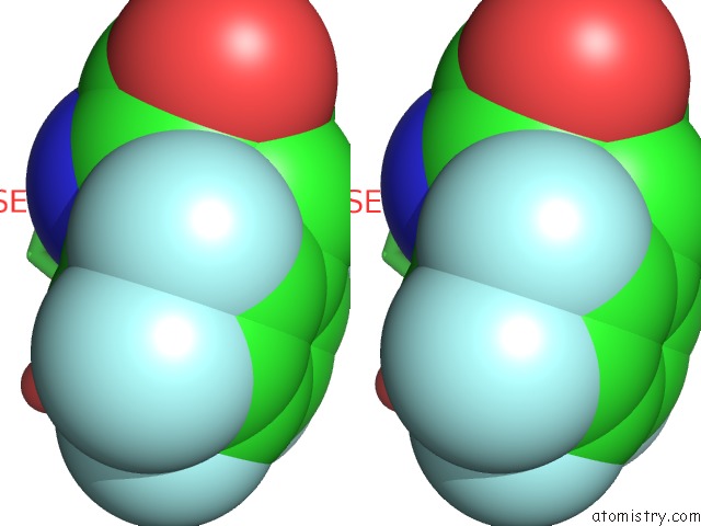
Fluorine binding site 2 out of 5 in 4rv3
Go back to
Fluorine binding site 2 out
of 5 in the Crystal Structure of A Pentafluoro-Phe Incorporated Phosphatidylinositol-Specific Phospholipase C (H258X)From Staphylococcus Aureus
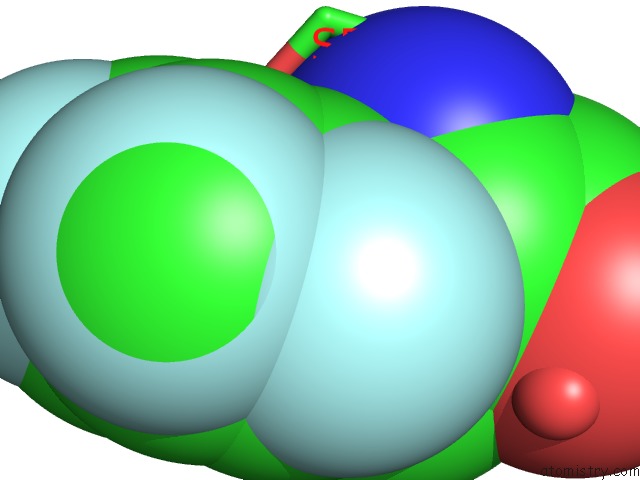
Mono view
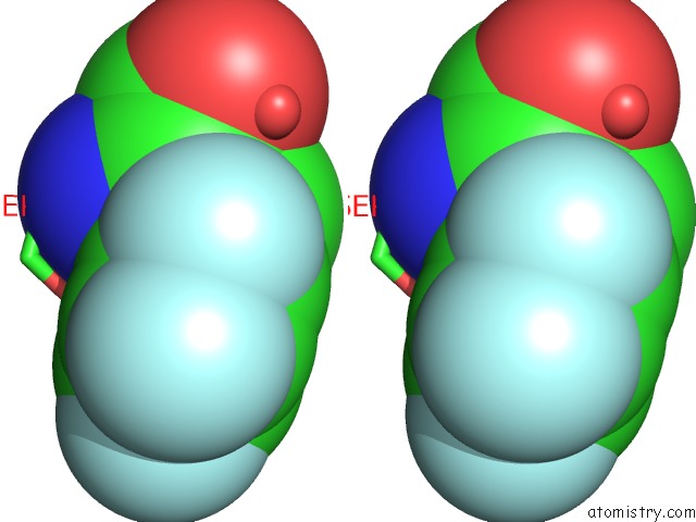
Stereo pair view
Mono view
Stereo pair view
A full contact list of Fluorine with other atoms in the F binding
site number 2 of Crystal Structure of A Pentafluoro-Phe Incorporated Phosphatidylinositol-Specific Phospholipase C (H258X)From Staphylococcus Aureus within 5.0Å range:
|
Fluorine binding site 3 out of 5 in 4rv3
Go back to
Fluorine binding site 3 out
of 5 in the Crystal Structure of A Pentafluoro-Phe Incorporated Phosphatidylinositol-Specific Phospholipase C (H258X)From Staphylococcus Aureus
Mono view
Stereo pair view
Mono view
Stereo pair view
A full contact list of Fluorine with other atoms in the F binding
site number 3 of Crystal Structure of A Pentafluoro-Phe Incorporated Phosphatidylinositol-Specific Phospholipase C (H258X)From Staphylococcus Aureus within 5.0Å range:
|
Fluorine binding site 4 out of 5 in 4rv3
Go back to
Fluorine binding site 4 out
of 5 in the Crystal Structure of A Pentafluoro-Phe Incorporated Phosphatidylinositol-Specific Phospholipase C (H258X)From Staphylococcus Aureus
Mono view
Stereo pair view
Mono view
Stereo pair view
A full contact list of Fluorine with other atoms in the F binding
site number 4 of Crystal Structure of A Pentafluoro-Phe Incorporated Phosphatidylinositol-Specific Phospholipase C (H258X)From Staphylococcus Aureus within 5.0Å range:
|
Fluorine binding site 5 out of 5 in 4rv3
Go back to
Fluorine binding site 5 out
of 5 in the Crystal Structure of A Pentafluoro-Phe Incorporated Phosphatidylinositol-Specific Phospholipase C (H258X)From Staphylococcus Aureus
Mono view
Stereo pair view
Mono view
Stereo pair view
A full contact list of Fluorine with other atoms in the F binding
site number 5 of Crystal Structure of A Pentafluoro-Phe Incorporated Phosphatidylinositol-Specific Phospholipase C (H258X)From Staphylococcus Aureus within 5.0Å range:
|
Reference:
T.He,
A.Gershenson,
S.J.Eyles,
Y.J.Lee,
W.R.Liu,
J.Wang,
J.Gao,
M.F.Roberts.
Fluorinated Aromatic Amino Acids Distinguish Cation-Pi Interactions From Membrane Insertion. J.Biol.Chem. V. 290 19334 2015.
ISSN: ISSN 0021-9258
PubMed: 26092728
DOI: 10.1074/JBC.M115.668343
Page generated: Tue Jul 15 00:41:41 2025
ISSN: ISSN 0021-9258
PubMed: 26092728
DOI: 10.1074/JBC.M115.668343
Last articles
Fe in 2YXOFe in 2YRS
Fe in 2YXC
Fe in 2YNM
Fe in 2YVJ
Fe in 2YP1
Fe in 2YU2
Fe in 2YU1
Fe in 2YQB
Fe in 2YOO