Fluorine »
PDB 4xyf-4ymo »
4y10 »
Fluorine in PDB 4y10: Trypsin in Complex with with Bpti Mutant (2S)-2-Amino-4,4- Difluorobutanoic Acid
Enzymatic activity of Trypsin in Complex with with Bpti Mutant (2S)-2-Amino-4,4- Difluorobutanoic Acid
All present enzymatic activity of Trypsin in Complex with with Bpti Mutant (2S)-2-Amino-4,4- Difluorobutanoic Acid:
3.4.21.4;
3.4.21.4;
Protein crystallography data
The structure of Trypsin in Complex with with Bpti Mutant (2S)-2-Amino-4,4- Difluorobutanoic Acid, PDB code: 4y10
was solved by
B.Loll,
S.Ye,
A.A.Berger,
U.Muelow,
C.Alings,
M.C.Wahl,
B.Koksch,
with X-Ray Crystallography technique. A brief refinement statistics is given in the table below:
| Resolution Low / High (Å) | 34.12 / 1.37 |
| Space group | I 2 2 2 |
| Cell size a, b, c (Å), α, β, γ (°) | 74.694, 81.854, 123.612, 90.00, 90.00, 90.00 |
| R / Rfree (%) | 13.8 / 16.5 |
Other elements in 4y10:
The structure of Trypsin in Complex with with Bpti Mutant (2S)-2-Amino-4,4- Difluorobutanoic Acid also contains other interesting chemical elements:
| Calcium | (Ca) | 1 atom |
Fluorine Binding Sites:
The binding sites of Fluorine atom in the Trypsin in Complex with with Bpti Mutant (2S)-2-Amino-4,4- Difluorobutanoic Acid
(pdb code 4y10). This binding sites where shown within
5.0 Angstroms radius around Fluorine atom.
In total 2 binding sites of Fluorine where determined in the Trypsin in Complex with with Bpti Mutant (2S)-2-Amino-4,4- Difluorobutanoic Acid, PDB code: 4y10:
Jump to Fluorine binding site number: 1; 2;
In total 2 binding sites of Fluorine where determined in the Trypsin in Complex with with Bpti Mutant (2S)-2-Amino-4,4- Difluorobutanoic Acid, PDB code: 4y10:
Jump to Fluorine binding site number: 1; 2;
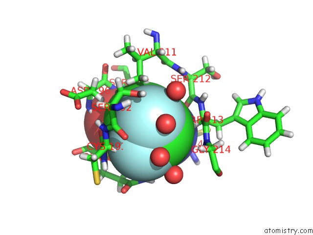
Fluorine binding site 1 out of 2 in 4y10
Go back to
Fluorine binding site 1 out
of 2 in the Trypsin in Complex with with Bpti Mutant (2S)-2-Amino-4,4- Difluorobutanoic Acid
Mono view

Stereo pair view
Mono view
Stereo pair view
A full contact list of Fluorine with other atoms in the F binding
site number 1 of Trypsin in Complex with with Bpti Mutant (2S)-2-Amino-4,4- Difluorobutanoic Acid within 5.0Å range:
|
Fluorine binding site 2 out of 2 in 4y10
Go back to
Fluorine binding site 2 out
of 2 in the Trypsin in Complex with with Bpti Mutant (2S)-2-Amino-4,4- Difluorobutanoic Acid

Mono view

Stereo pair view
Mono view
Stereo pair view
A full contact list of Fluorine with other atoms in the F binding
site number 2 of Trypsin in Complex with with Bpti Mutant (2S)-2-Amino-4,4- Difluorobutanoic Acid within 5.0Å range:
|
Reference:
S.Ye,
B.Loll,
A.A.Berger,
U.Mulow,
C.Alings,
M.C.Wahl,
B.Koksch.
Fluorine Teams Up with Water to Restore Inhibitor Activity to Mutant Bpti. Chem Sci V. 6 5246 2015.
ISSN: ISSN 2041-6520
PubMed: 29449928
DOI: 10.1039/C4SC03227F
Page generated: Thu Aug 1 06:52:57 2024
ISSN: ISSN 2041-6520
PubMed: 29449928
DOI: 10.1039/C4SC03227F
Last articles
Zn in 9MJ5Zn in 9HNW
Zn in 9G0L
Zn in 9FNE
Zn in 9DZN
Zn in 9E0I
Zn in 9D32
Zn in 9DAK
Zn in 8ZXC
Zn in 8ZUF