Fluorine »
PDB 4xyf-4ymo »
4y2q »
Fluorine in PDB 4y2q: Structure of Soluble Epoxide Hydrolase in Complex with 1-[3- (Trifluoromethyl)Pyridin-2-Yl]Piperazine
Enzymatic activity of Structure of Soluble Epoxide Hydrolase in Complex with 1-[3- (Trifluoromethyl)Pyridin-2-Yl]Piperazine
All present enzymatic activity of Structure of Soluble Epoxide Hydrolase in Complex with 1-[3- (Trifluoromethyl)Pyridin-2-Yl]Piperazine:
3.3.2.10;
3.3.2.10;
Protein crystallography data
The structure of Structure of Soluble Epoxide Hydrolase in Complex with 1-[3- (Trifluoromethyl)Pyridin-2-Yl]Piperazine, PDB code: 4y2q
was solved by
Y.Amano,
T.Yamaguchi,
with X-Ray Crystallography technique. A brief refinement statistics is given in the table below:
| Resolution Low / High (Å) | 76.13 / 2.40 |
| Space group | P 65 2 2 |
| Cell size a, b, c (Å), α, β, γ (°) | 92.544, 92.544, 243.634, 90.00, 90.00, 120.00 |
| R / Rfree (%) | 19.6 / 25.7 |
Other elements in 4y2q:
The structure of Structure of Soluble Epoxide Hydrolase in Complex with 1-[3- (Trifluoromethyl)Pyridin-2-Yl]Piperazine also contains other interesting chemical elements:
| Magnesium | (Mg) | 1 atom |
Fluorine Binding Sites:
The binding sites of Fluorine atom in the Structure of Soluble Epoxide Hydrolase in Complex with 1-[3- (Trifluoromethyl)Pyridin-2-Yl]Piperazine
(pdb code 4y2q). This binding sites where shown within
5.0 Angstroms radius around Fluorine atom.
In total 3 binding sites of Fluorine where determined in the Structure of Soluble Epoxide Hydrolase in Complex with 1-[3- (Trifluoromethyl)Pyridin-2-Yl]Piperazine, PDB code: 4y2q:
Jump to Fluorine binding site number: 1; 2; 3;
In total 3 binding sites of Fluorine where determined in the Structure of Soluble Epoxide Hydrolase in Complex with 1-[3- (Trifluoromethyl)Pyridin-2-Yl]Piperazine, PDB code: 4y2q:
Jump to Fluorine binding site number: 1; 2; 3;
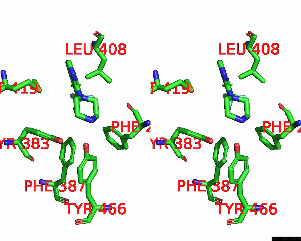
Fluorine binding site 1 out of 3 in 4y2q
Go back to
Fluorine binding site 1 out
of 3 in the Structure of Soluble Epoxide Hydrolase in Complex with 1-[3- (Trifluoromethyl)Pyridin-2-Yl]Piperazine

Mono view

Stereo pair view
Mono view
Stereo pair view
A full contact list of Fluorine with other atoms in the F binding
site number 1 of Structure of Soluble Epoxide Hydrolase in Complex with 1-[3- (Trifluoromethyl)Pyridin-2-Yl]Piperazine within 5.0Å range:
|
Fluorine binding site 2 out of 3 in 4y2q
Go back to
Fluorine binding site 2 out
of 3 in the Structure of Soluble Epoxide Hydrolase in Complex with 1-[3- (Trifluoromethyl)Pyridin-2-Yl]Piperazine

Mono view

Stereo pair view
Mono view
Stereo pair view
A full contact list of Fluorine with other atoms in the F binding
site number 2 of Structure of Soluble Epoxide Hydrolase in Complex with 1-[3- (Trifluoromethyl)Pyridin-2-Yl]Piperazine within 5.0Å range:
|
Fluorine binding site 3 out of 3 in 4y2q
Go back to
Fluorine binding site 3 out
of 3 in the Structure of Soluble Epoxide Hydrolase in Complex with 1-[3- (Trifluoromethyl)Pyridin-2-Yl]Piperazine

Mono view
Stereo pair view
Mono view
Stereo pair view
A full contact list of Fluorine with other atoms in the F binding
site number 3 of Structure of Soluble Epoxide Hydrolase in Complex with 1-[3- (Trifluoromethyl)Pyridin-2-Yl]Piperazine within 5.0Å range:
|
Reference:
Y.Amano,
E.Tanabe,
T.Yamaguchi.
Identification of N-Ethylmethylamine As A Novel Scaffold For Inhibitors of Soluble Epoxide Hydrolase By Crystallographic Fragment Screening Bioorg.Med.Chem. V. 23 2310 2015.
ISSN: ESSN 1464-3391
PubMed: 25862210
DOI: 10.1016/J.BMC.2015.03.083
Page generated: Thu Aug 1 06:52:57 2024
ISSN: ESSN 1464-3391
PubMed: 25862210
DOI: 10.1016/J.BMC.2015.03.083
Last articles
Zn in 9JYWZn in 9IR4
Zn in 9IR3
Zn in 9GMX
Zn in 9GMW
Zn in 9JEJ
Zn in 9ERF
Zn in 9ERE
Zn in 9EGV
Zn in 9EGW