Fluorine »
PDB 4zga-4zzi »
4zy2 »
Fluorine in PDB 4zy2: X-Ray Crystal Structure of Pfa-M17 in Complex with Hydroxamic Acid- Based Inhibitor 10O
Protein crystallography data
The structure of X-Ray Crystal Structure of Pfa-M17 in Complex with Hydroxamic Acid- Based Inhibitor 10O, PDB code: 4zy2
was solved by
N.Drinkwater,
S.Mcgowan,
with X-Ray Crystallography technique. A brief refinement statistics is given in the table below:
| Resolution Low / High (Å) | 48.54 / 2.10 |
| Space group | P 21 21 21 |
| Cell size a, b, c (Å), α, β, γ (°) | 174.060, 177.247, 230.773, 90.00, 90.00, 90.00 |
| R / Rfree (%) | 19.4 / 23.9 |
Other elements in 4zy2:
The structure of X-Ray Crystal Structure of Pfa-M17 in Complex with Hydroxamic Acid- Based Inhibitor 10O also contains other interesting chemical elements:
| Zinc | (Zn) | 24 atoms |
Fluorine Binding Sites:
Pages:
>>> Page 1 <<< Page 2, Binding sites: 11 - 20; Page 3, Binding sites: 21 - 30; Page 4, Binding sites: 31 - 36;Binding sites:
The binding sites of Fluorine atom in the X-Ray Crystal Structure of Pfa-M17 in Complex with Hydroxamic Acid- Based Inhibitor 10O (pdb code 4zy2). This binding sites where shown within 5.0 Angstroms radius around Fluorine atom.In total 36 binding sites of Fluorine where determined in the X-Ray Crystal Structure of Pfa-M17 in Complex with Hydroxamic Acid- Based Inhibitor 10O, PDB code: 4zy2:
Jump to Fluorine binding site number: 1; 2; 3; 4; 5; 6; 7; 8; 9; 10;
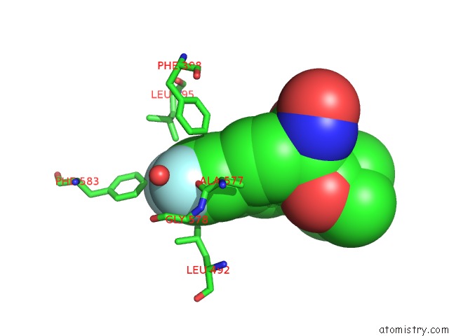
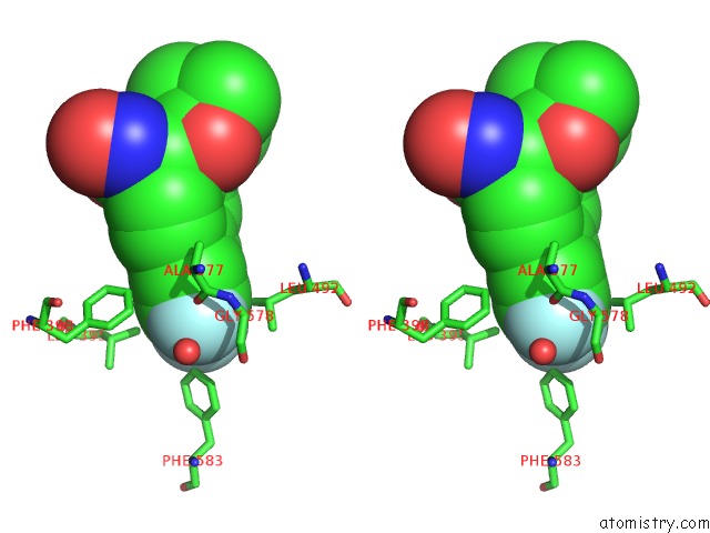
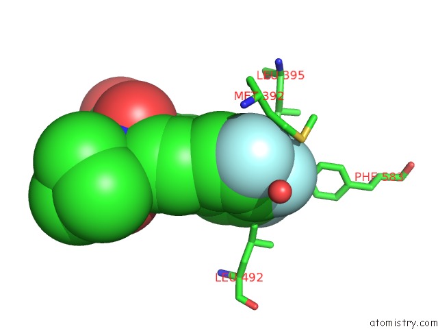
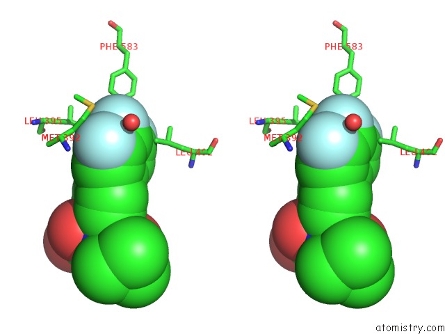
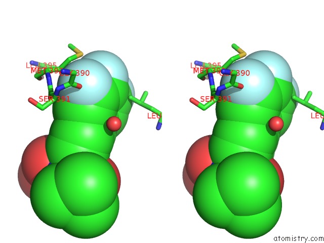
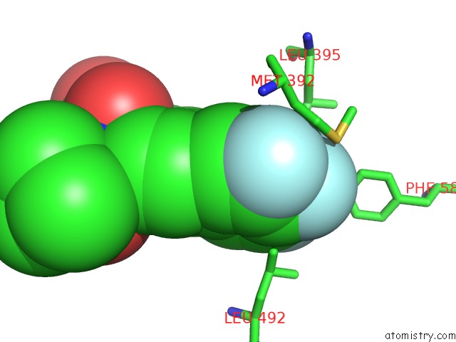
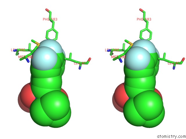
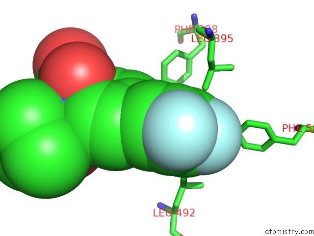
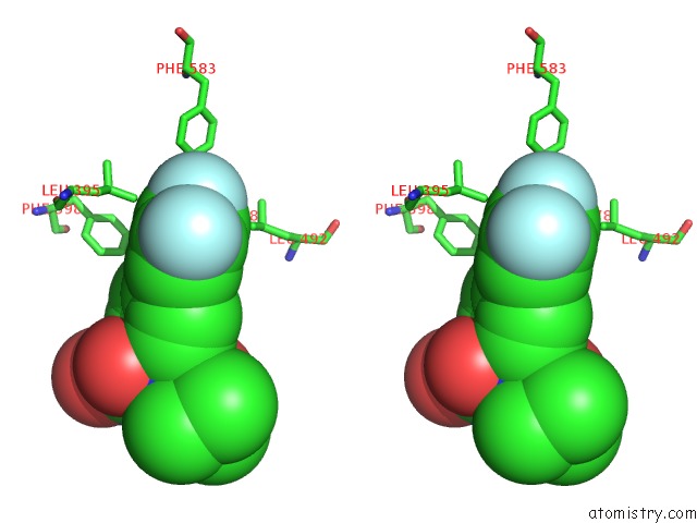
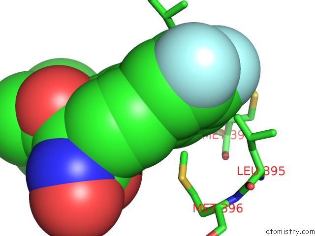
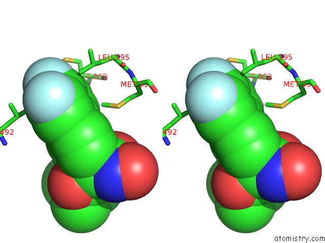
Fluorine binding site 1 out of 36 in 4zy2
Go back to
Fluorine binding site 1 out
of 36 in the X-Ray Crystal Structure of Pfa-M17 in Complex with Hydroxamic Acid- Based Inhibitor 10O
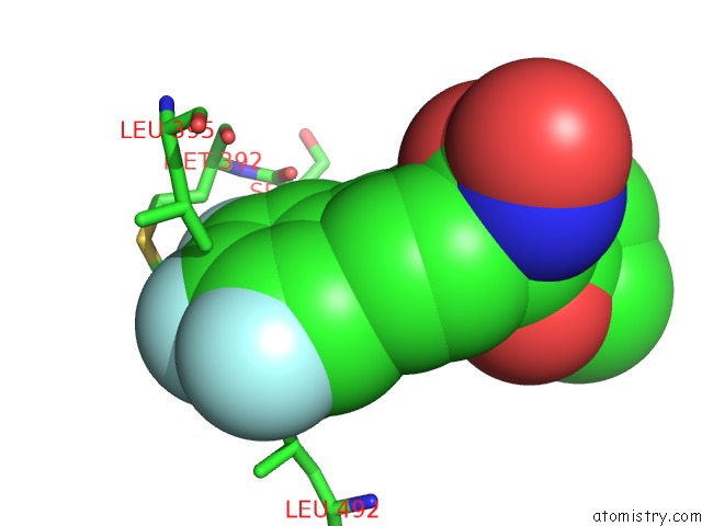
Mono view
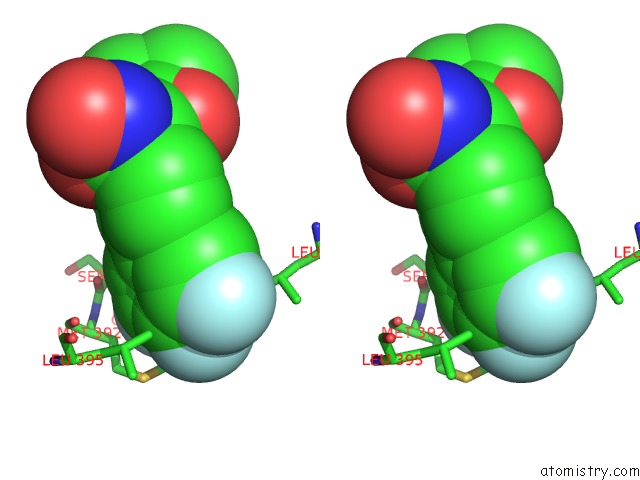
Stereo pair view
Mono view
Stereo pair view
A full contact list of Fluorine with other atoms in the F binding
site number 1 of X-Ray Crystal Structure of Pfa-M17 in Complex with Hydroxamic Acid- Based Inhibitor 10O within 5.0Å range:
|
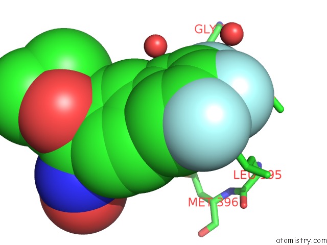
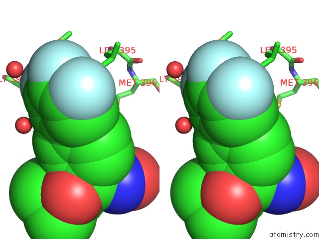
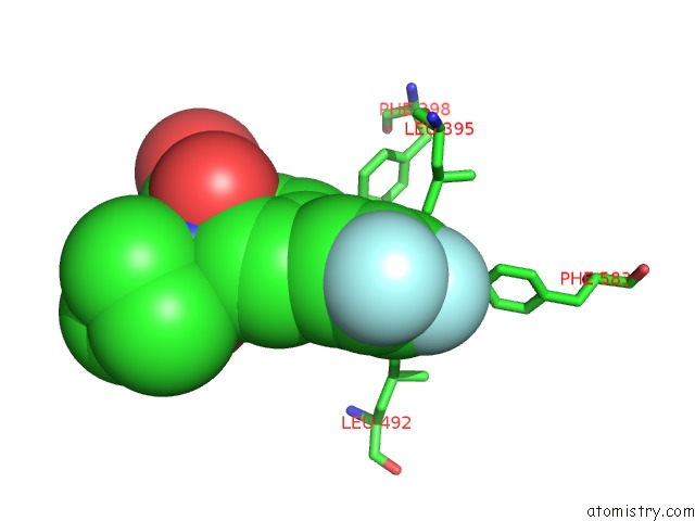
Fluorine binding site 2 out of 36 in 4zy2
Go back to
Fluorine binding site 2 out
of 36 in the X-Ray Crystal Structure of Pfa-M17 in Complex with Hydroxamic Acid- Based Inhibitor 10O
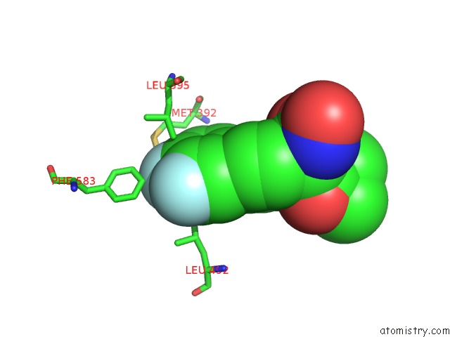
Mono view
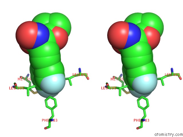
Stereo pair view
Mono view
Stereo pair view
A full contact list of Fluorine with other atoms in the F binding
site number 2 of X-Ray Crystal Structure of Pfa-M17 in Complex with Hydroxamic Acid- Based Inhibitor 10O within 5.0Å range:
|
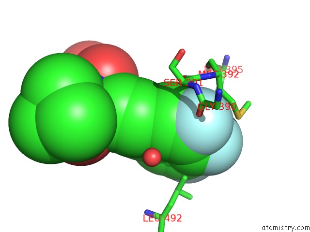
Fluorine binding site 3 out of 36 in 4zy2
Go back to
Fluorine binding site 3 out
of 36 in the X-Ray Crystal Structure of Pfa-M17 in Complex with Hydroxamic Acid- Based Inhibitor 10O
Mono view
Stereo pair view
Mono view
Stereo pair view
A full contact list of Fluorine with other atoms in the F binding
site number 3 of X-Ray Crystal Structure of Pfa-M17 in Complex with Hydroxamic Acid- Based Inhibitor 10O within 5.0Å range:
|
Fluorine binding site 4 out of 36 in 4zy2
Go back to
Fluorine binding site 4 out
of 36 in the X-Ray Crystal Structure of Pfa-M17 in Complex with Hydroxamic Acid- Based Inhibitor 10O
Mono view
Stereo pair view
Mono view
Stereo pair view
A full contact list of Fluorine with other atoms in the F binding
site number 4 of X-Ray Crystal Structure of Pfa-M17 in Complex with Hydroxamic Acid- Based Inhibitor 10O within 5.0Å range:
|
Fluorine binding site 5 out of 36 in 4zy2
Go back to
Fluorine binding site 5 out
of 36 in the X-Ray Crystal Structure of Pfa-M17 in Complex with Hydroxamic Acid- Based Inhibitor 10O
Mono view
Stereo pair view
Mono view
Stereo pair view
A full contact list of Fluorine with other atoms in the F binding
site number 5 of X-Ray Crystal Structure of Pfa-M17 in Complex with Hydroxamic Acid- Based Inhibitor 10O within 5.0Å range:
|
Fluorine binding site 6 out of 36 in 4zy2
Go back to
Fluorine binding site 6 out
of 36 in the X-Ray Crystal Structure of Pfa-M17 in Complex with Hydroxamic Acid- Based Inhibitor 10O
Mono view

Stereo pair view
Mono view
Stereo pair view
A full contact list of Fluorine with other atoms in the F binding
site number 6 of X-Ray Crystal Structure of Pfa-M17 in Complex with Hydroxamic Acid- Based Inhibitor 10O within 5.0Å range:
|
Fluorine binding site 7 out of 36 in 4zy2
Go back to
Fluorine binding site 7 out
of 36 in the X-Ray Crystal Structure of Pfa-M17 in Complex with Hydroxamic Acid- Based Inhibitor 10O
Mono view
Stereo pair view
Mono view
Stereo pair view
A full contact list of Fluorine with other atoms in the F binding
site number 7 of X-Ray Crystal Structure of Pfa-M17 in Complex with Hydroxamic Acid- Based Inhibitor 10O within 5.0Å range:
|
Fluorine binding site 8 out of 36 in 4zy2
Go back to
Fluorine binding site 8 out
of 36 in the X-Ray Crystal Structure of Pfa-M17 in Complex with Hydroxamic Acid- Based Inhibitor 10O
Mono view
Stereo pair view
Mono view
Stereo pair view
A full contact list of Fluorine with other atoms in the F binding
site number 8 of X-Ray Crystal Structure of Pfa-M17 in Complex with Hydroxamic Acid- Based Inhibitor 10O within 5.0Å range:
|
Fluorine binding site 9 out of 36 in 4zy2
Go back to
Fluorine binding site 9 out
of 36 in the X-Ray Crystal Structure of Pfa-M17 in Complex with Hydroxamic Acid- Based Inhibitor 10O
Mono view
Stereo pair view
Mono view
Stereo pair view
A full contact list of Fluorine with other atoms in the F binding
site number 9 of X-Ray Crystal Structure of Pfa-M17 in Complex with Hydroxamic Acid- Based Inhibitor 10O within 5.0Å range:
|
Fluorine binding site 10 out of 36 in 4zy2
Go back to
Fluorine binding site 10 out
of 36 in the X-Ray Crystal Structure of Pfa-M17 in Complex with Hydroxamic Acid- Based Inhibitor 10O
Mono view
Stereo pair view
Mono view
Stereo pair view
A full contact list of Fluorine with other atoms in the F binding
site number 10 of X-Ray Crystal Structure of Pfa-M17 in Complex with Hydroxamic Acid- Based Inhibitor 10O within 5.0Å range:
|
Reference:
N.Drinkwater,
N.B.Vinh,
S.N.Mistry,
R.S.Bamert,
C.Ruggeri,
J.P.Holleran,
S.Loganathan,
A.Paiardini,
S.A.Charman,
A.K.Powell,
V.M.Avery,
S.Mcgowan,
P.J.Scammells.
Potent Dual Inhibitors of Plasmodium Falciparum M1 and M17 Aminopeptidases Through Optimization of S1 Pocket Interactions. Eur.J.Med.Chem. V. 110 43 2016.
ISSN: ISSN 0223-5234
PubMed: 26807544
DOI: 10.1016/J.EJMECH.2016.01.015
Page generated: Thu Aug 1 07:28:10 2024
ISSN: ISSN 0223-5234
PubMed: 26807544
DOI: 10.1016/J.EJMECH.2016.01.015
Last articles
F in 4DSAF in 4DQ5
F in 4DPU
F in 4DPT
F in 4DOA
F in 4DMY
F in 4DO9
F in 4DLC
F in 4DMX
F in 4DL8