Fluorine »
PDB 4zzj-5ah8 »
5aeg »
Fluorine in PDB 5aeg: A Bacterial Protein Structure in Glycoside Hydrolase Family 31.
Enzymatic activity of A Bacterial Protein Structure in Glycoside Hydrolase Family 31.
All present enzymatic activity of A Bacterial Protein Structure in Glycoside Hydrolase Family 31.:
3.2.1.20;
3.2.1.20;
Protein crystallography data
The structure of A Bacterial Protein Structure in Glycoside Hydrolase Family 31., PDB code: 5aeg
was solved by
Y.Jin,
G.Speciale,
G.J.Davies,
S.J.Williams,
E.D.Goddard-Borger,
with X-Ray Crystallography technique. A brief refinement statistics is given in the table below:
| Resolution Low / High (Å) | 105.45 / 1.85 |
| Space group | P 1 21 1 |
| Cell size a, b, c (Å), α, β, γ (°) | 78.665, 112.643, 111.881, 90.00, 109.52, 90.00 |
| R / Rfree (%) | 16 / 19.3 |
Other elements in 5aeg:
The structure of A Bacterial Protein Structure in Glycoside Hydrolase Family 31. also contains other interesting chemical elements:
| Chlorine | (Cl) | 2 atoms |
| Calcium | (Ca) | 3 atoms |
Fluorine Binding Sites:
The binding sites of Fluorine atom in the A Bacterial Protein Structure in Glycoside Hydrolase Family 31.
(pdb code 5aeg). This binding sites where shown within
5.0 Angstroms radius around Fluorine atom.
In total only one binding site of Fluorine was determined in the A Bacterial Protein Structure in Glycoside Hydrolase Family 31., PDB code: 5aeg:
In total only one binding site of Fluorine was determined in the A Bacterial Protein Structure in Glycoside Hydrolase Family 31., PDB code: 5aeg:
Fluorine binding site 1 out of 1 in 5aeg
Go back to
Fluorine binding site 1 out
of 1 in the A Bacterial Protein Structure in Glycoside Hydrolase Family 31.
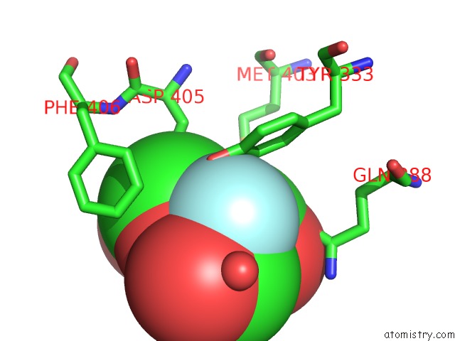
Mono view
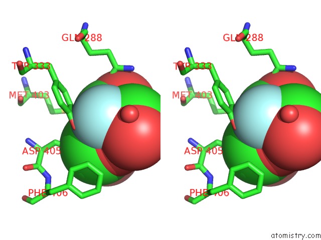
Stereo pair view
Mono view
Stereo pair view
A full contact list of Fluorine with other atoms in the F binding
site number 1 of A Bacterial Protein Structure in Glycoside Hydrolase Family 31. within 5.0Å range:
|
Reference:
G.Speciale,
Y.Jin,
G.J.Davies,
S.J.Williams,
E.D.Goddard-Borger.
Yihq Is A Sulfoquinovosidase That Cleaves Sulfoquinovosyl Diacylglyceride Sulfolipids. Nat.Chem.Biol. V. 12 215 2016.
ISSN: ISSN 1552-4450
PubMed: 26878550
DOI: 10.1038/NCHEMBIO.2023
Page generated: Thu Aug 1 07:41:23 2024
ISSN: ISSN 1552-4450
PubMed: 26878550
DOI: 10.1038/NCHEMBIO.2023
Last articles
Zn in 9J0NZn in 9J0O
Zn in 9J0P
Zn in 9FJX
Zn in 9EKB
Zn in 9C0F
Zn in 9CAH
Zn in 9CH0
Zn in 9CH3
Zn in 9CH1